When you hear the word “orbit,” you likely picture planets orbiting the sun, or think of a minty fresh gum, but the term “orbit” also refers to the bones, muscles, ligaments, fat, and other tissues that make up the eye socket. A variety of diseases can affect your pet’s or your horse’s orbit—here are the five most common:
#1: Orbital cellulitis and abscesses in pets and horses
The orbital contents can become inflamed or infected (i.e., orbital cellulitis) for several reasons:
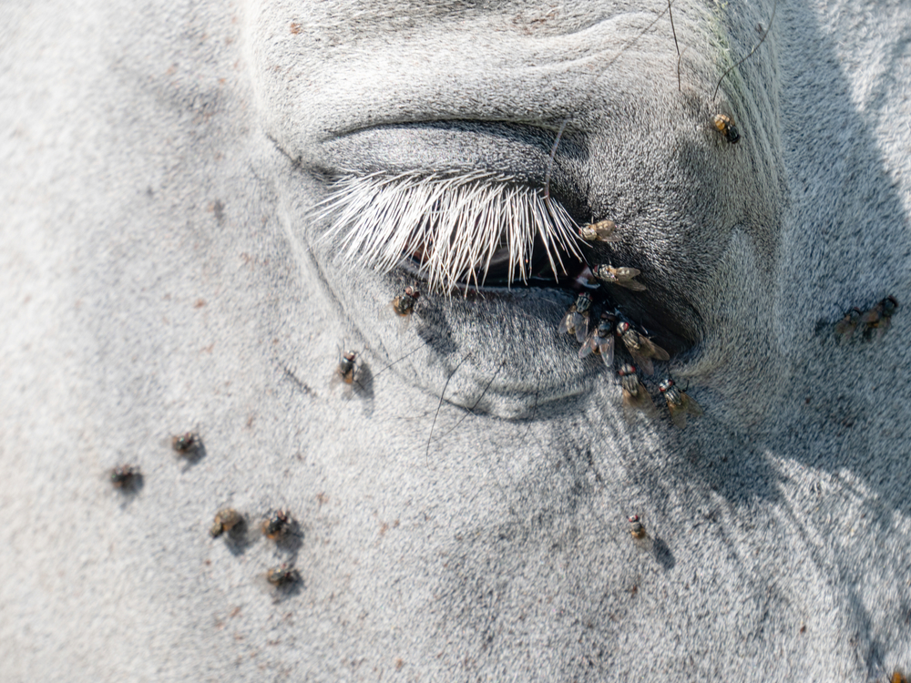
- Foreign material — Grass awns, porcupine quills, wood slivers, or other foreign materials may lodge in the orbit.
- Infection from other areas — Infectious agents can enter the orbit from the bloodstream or sinuses. Anatomically in dogs and cats, the back portion of the gums and upper tooth roots are close to the bottom of the orbit, so mouth infections can also easily extend into the orbit.
- Trauma — Blunt force trauma to the eye or side of the face can cause orbital inflammation or create a hematoma (i.e., an area of free blood in tissue), which bacteria then invade.
Orbital cellulitis is sometimes accompanied by a discrete pocket of purulent material (i.e., an orbital abscess) that may or may not contain a foreign object. Affected animals typically experience rapid onset of an inflamed, bulging eye, abnormal eye discharge, elevated third eyelid, fever, and pain when opening their mouth. Treatment involves antibiotics, anti-inflammatory drugs, and draining any abscess. Generally, prognosis is good, and the eye appears more normal three to four days after starting treatment.
#2: Orbital masses in pets and horses
Older animals are at risk for tumor formation in the orbit, which may affect vision if the optic nerve is damaged. The affected eye may appear pushed forward, and is usually non-painful initially. Approximately 80% to 90% of pet orbital tumors are cancerous, with meningioma, fibroma, osteosarcoma, or lymphoma the most common. Horses typically have squamous cell carcinoma, lymphoma, or tumor invasion from adjacent locations. The tumor can be removed without affecting the eye in some cases, but in others, removal of the eye and the orbit’s contents may be necessary. Adjunctive therapy, such as chemotherapy or radiation, may also be considered, but overall prognosis is usually poor regardless of treatment approach.
#3: Orbital trauma in horses and pets
Horses are prone to fracturing the orbit’s outer bony rim, which protrudes further from the side of their face than other structures. Orbital fractures may occur when the horse is kicked, rears in a confined location, or runs into an object, and require medical and surgical management for the best prognosis. Trauma can also cause orbital fractures in pets, but because their orbit has a different conformation, pets are also prone to proptosis, a condition where the eyeball pops out of the socket and rests in front of the eyelids. This condition occurs more commonly in brachycephalic (i.e., short-faced) animals, because their eye sockets tend to be shallow. Proptosed eyes must be surgically replaced into the socket under general anesthesia as soon as possible for the best chance of a good outcome. About 40% of dogs with proptosis, and almost no cats, will regain vision, and significantly damaged eyes may require removal.
#4: Myositis in dogs
Some dog breeds are predisposed to myositis (i.e., muscle inflammation) in the muscles surrounding the eye, or those used for chewing. The resulting muscle swelling compresses the orbit and both eyes appear pushed forward. Affected dogs may also have pain and difficulty opening their mouth. Immunosuppressive drugs are used for myositis management, but relapses are common, unfortunately, and can cause long-term damage.
#5: Orbital cysts in pets
Orbital cysts, which are less common than other orbital problems, can be congenital or trauma-related. When the salivary gland is traumatized, saliva may leak out, create a fluid pocket, and cause inflammation that manifests as swelling, third eyelid elevation, and a bulging eye. Surgery is typically required to address orbital cysts.
Orbital disease diagnostics

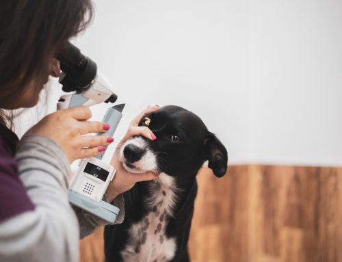
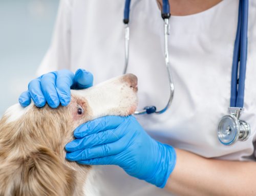
Dr. Pierce will always perform a thorough ophthalmic exam, including palpating the eyes and orbits, and measuring intraocular pressure. He will also closely examine your pet’s mouth, teeth, gums, nose, and sinuses for any abnormalities or infection indicators. Sometimes he will use imaging, such as an eye ultrasound, X-ray, CT, or MRI, to further evaluate the orbit and reach a final diagnosis. Additional disease-specific diagnostics, such as a muscle biopsy for myositis, culture and cytology (i.e., cell examination) for orbital abscesses or cellulitis, or a fine needle aspirate (FNA) or biopsy for suspected tumors, may be useful.
If you or your family veterinarian suspects your pet or horse could have an orbital disease, our Veterinary Vision Center team is here to give them the care they deserve. Contact us to set up an appointment with Dr. Pierce, our superb board-certified veterinary ophthalmologist.

Leave A Comment