
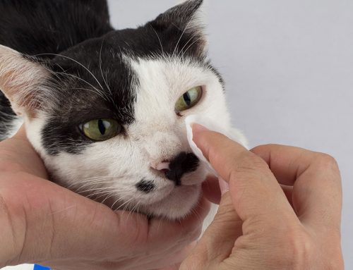
Numerous conditions can result in a dysfunction called Horner’s syndrome, which is a neurological disorder that affects the nerves innervating a dog’s eye and facial muscles. Our team at Veterinary Vision Center wants to provide information about this syndrome, in case your dog is affected.
Horner’s syndrome signs in dogs
A syndrome is a condition characterized by a collection of signs, and Horner’s syndrome signs are caused by damage to the sympathetic nervous system, whose nerves supply the eye and facial muscles on the affected side. This results in:
- Constricted pupil
- Elevated third eyelid
- Eyeball retraction (i.e., the eye appears shrunken)
- Eyelid drooping
- Increased pink color, and nose and ear warmth on the affected side
Understanding the dog’s sympathetic nervous system
The nervous system is composed of the somatic nervous system, which supplies skin and muscles and is involved in conscious activity, and the autonomic nervous system, which controls bodily functions not consciously directed, such as heart and respiratory rates, blood circulation, and pupil dilation and constriction. The autonomic nervous system is composed of two antagonistic nerve sets:
- Parasympathetic nervous system — The parasympathetic nervous system maintains the body during a normal state. In the eye region, this system constricts the pupil, raises the third eyelid, and retracts the eye for protection.
- Sympathetic nervous system — The sympathetic nervous system prepares the body for a fight or flight situation. In the eye region, this system dilates the pupil, widens the eyelids, drops the third eyelid, and keeps the eye in a forward position in the socket.
These two nervous systems work together to maintain balance in a healthy body, with one system slightly dominating the other, depending on the situation. When the sympathetic nerves supplying the eye region are damaged, only the parasympathetic nerves work, causing Horner’s syndrome.
Damage to the dog’s sympathetic nervous system
The sympathetic nerves that innervate the eye region can be damaged at any point on their long path from the brain. Nerve segments include:
- Central segment — The first segment begins as the nerves exit the brain at the hypothalamus and travel through the brainstem and down the spinal cord in the neck area. Damage in this area is called a first order lesion, and includes brain tumors or trauma, vascular brain accidents, and herniated vertebral discs.
- Preganglionic segment — The nerves exit the spinal cord inside the chest at the the second thoracic vertebra level, and travel on the right and left sides up the neck to the middle ear vicinity. Preganglionic segment damage is called a second order lesion, and can occur when the foreleg is pulled and the armpit area nerves are overstretched, from neck trauma, such as pulling extremely hard on a leash, and neck or chest tumors.
- Postganglionic segment — The last segment, the postganglionic segment, starts below the ear and travels to the eye. Postganglionic segment damage is called a third order lesion and involves infection or trauma to the middle ear. These lesions are the most common cause of Horner’s syndrome in dogs.
Localizing the lesion causing the dog’s Horner’s syndrome
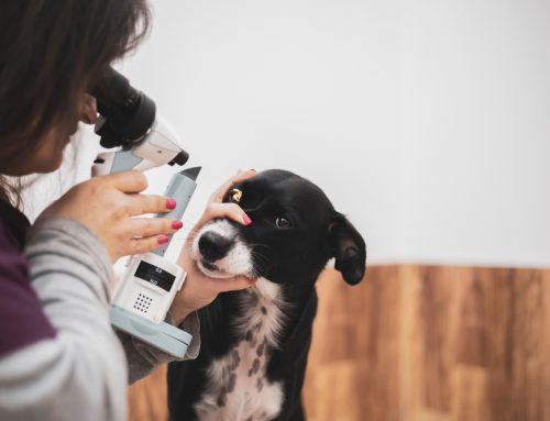
When an ear infection, injury, or tumor isn’t apparent, the lesion must be localized to determine the underlying cause. Medications, such as specific eye drops, can be administered to stimulate different sympathetic nervous system segments to help determine the damaged area, and imaging modalities, including X-rays, magnetic resonance imaging (MRI), and computed tomography (CT scan), may also be used. When the lesion cannot be determined, Horner’s syndrome is classified as idiopathic. Golden retrievers are at higher risk for the idiopathic condition.
Treating Horner’s syndrome in dogs

Horner’s syndrome is not painful and does not affect the dog’s vision, so treatment isn’t necessary. However, Horner’s syndrome indicates underlying nerve damage, and the cause may need addressing. When the cause can be determined, your veterinarian will develop an appropriate treatment plan to address the condition and alleviate your dog’s signs. When the cause is not known, natural recovery usually occurs in 16 weeks to 6 months, depending on sign severity.
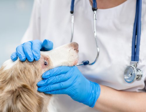
Any neurological issue affecting your dog is concerning, and a pet who is exhibiting Horner’s syndrome signs must be evaluated by a veterinary professional to determine the cause. Contact our team at Veterinary Vision Center if your dog has a constricted pupil, drooping eyelid, retracted eyeball, or elevated third eyelid, so we can localize the lesion and develop an appropriate treatment plan.

Leave A Comment