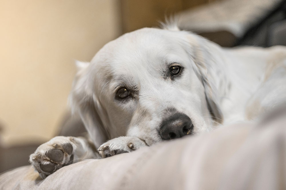
Puppies and kittens are born with their eyes closed, and over the first few weeks of life, their visual system develops rapidly. Most of the time, this process unfolds exactly as it should. But some young animals are born with congenital eye conditions (structural abnormalities present at birth) or develop hereditary eye diseases (conditions passed down genetically that may not become apparent until weeks or months later) that can affect their vision, comfort, or both if not caught early.
The earlier these conditions are identified, the better the chances of preserving vision and preventing complications. At Veterinary Vision Center, Dr. Pierce (a board-certified veterinary ophthalmologist) evaluates puppies and kittens referred by primary care veterinarians for eye concerns ranging from cloudiness and discharge to structural abnormalities that need specialist-level assessment. Our ophthalmic diagnostic services are designed to detect conditions that a standard wellness exam may not reveal. If your veterinarian has recommended an eye specialist evaluation for your young pet, call us at (318) 797-5522 or request an appointment to schedule a consultation.
Normal Eye Development: What to Expect
The Timeline of Eye Opening
Puppies and kittens are born with their eyelids fused shut as a protective measure. Eye opening typically occurs between 10 and 14 days of age, though the exact timing varies by individual. Vision development proceeds in stages: even after the eyes open, visual acuity is limited, and the system continues to mature through the first weeks and months of life. Eye color in young animals often shifts during this period and should stabilize by 12 weeks.
Visual development during early kitten and puppyhood is experience-dependent. The brain requires light and visual input through the eyes to wire the neural pathways that produce normal vision. This is one reason why conditions that block light from reaching the retina, such as dense congenital cataracts, need intervention early to preserve functional vision.
Key Structures and How They Work
Eye structure and function in dogs and cats centers on several components working together:
- Cornea: the clear outer surface that refracts light
- Lens: focuses light onto the retina
- Retina: the light-sensitive tissue layer at the back of the eye where photoreceptors convert light to signals
- Optic nerve: transmits those signals to the brain
Disruption at any point along this pathway, whether from structural abnormality, disease, or injury, can affect the clarity and quality of vision. Knowing what looks normal helps you recognize when something looks or behaves differently.
Congenital Eye Conditions
Present at Birth
Congenital eye defects are abnormalities of eye development present from birth. Some are identified at the first veterinary examination; others become apparent as the puppy or kitten begins to navigate visually.
- Microphthalmia: abnormally small eye development, which may involve structural disorganization within the eye. Severity ranges from mild and functionally acceptable to complete absence of usable vision in the affected eye.
- Persistent pupillary membranes (PPM): remnants of fetal tissue that normally regresses before birth remaining across the pupil. Minor PPM often have no functional significance; extensive strands can interfere with vision.
- Persistent hyperplastic primary vitreous (PHPV): failure of the embryonic blood vessel system in the eye to regress properly, leaving opaque material in or around the lens.
- Dermoids: skin-like tissue in an abnormal ocular location, typically at the corneal surface, causing irritation and discharge.
Hereditary Conditions That Develop Later
Hereditary eye disease encompasses a range of genetically transmitted conditions that may not be visible at birth but develop during growth or adulthood.
- Juvenile hereditary cataracts: lens opacification that begins in young dogs and progresses to significant visual impairment. Common in Labrador Retrievers, Golden Retrievers, Staffordshire Bull Terriers, and other breeds. Genetic testing of breeding animals reduces incidence.
- Progressive retinal atrophy (PRA): gradual degeneration of the retinal photoreceptors leading to progressive vision loss. Affects multiple dog and cat breeds. Night vision is often affected first. Genetic testing is available for many breed-specific mutations.
- Collie Eye Anomaly (CEA): a spectrum of hereditary developmental abnormalities of the choroid and sclera in Collies, Border Collies, Shetland Sheepdogs, and related breeds. Mild cases may not affect vision; severe cases involve retinal detachment.
- Hereditary cataracts in specific breeds: many breeds carry breed-specific mutations. Responsible breeders use health testing to screen breeding stock.

Eyelid and Structural Abnormalities
Entropion and Ectropion in Young Pets
Eyelid conformation problems are common in puppies of specific breeds and can begin causing corneal damage before the pet is fully grown.
Entropion in puppies is the inward rolling of the eyelid margin, pressing fur and lashes against the cornea with every blink. Constant friction causes corneal ulceration, pain, and over time, scarring that permanently affects vision. In very young puppies, temporary tacking sutures can hold the eyelid in the correct position while the face develops. Definitive surgical correction is performed once growth is complete.
Ectropion is the outward sagging of the lower lid, exposing conjunctival tissue and creating a reservoir for debris and bacteria. Chronic irritation and recurring infections follow.
Both conditions are corrected surgically once the diagnosis is confirmed and the patient’s growth is appropriately advanced.
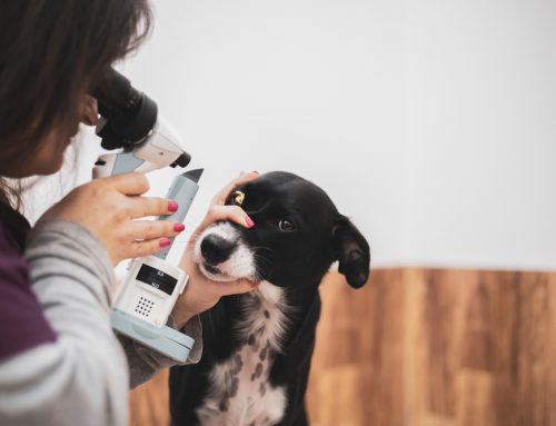
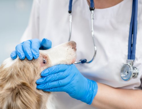
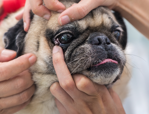
What a Pediatric Eye Exam Includes
Physical examination of the eye by a veterinary ophthalmologist goes significantly beyond the brief ocular check performed at a routine wellness visit.
A specialist evaluation includes:
- External inspection of lid margin position, lash direction, and periocular anatomy
- Biomicroscopy (slit lamp) examination of the cornea, anterior chamber, and lens
- Indirect ophthalmoscopy of the retina and optic nerve
- Tonometry to measure intraocular pressure
- Fluorescein stain to detect corneal ulcers
- Schirmer tear test for tear production
- Electroretinography when retinal function assessment is indicated
This level of evaluation detects subtle changes and pre-clinical disease that would not be apparent on a general physical examination.
Conditions That Develop During Growth
Juvenile Glaucoma
Canine glaucoma includes both primary hereditary forms and secondary forms arising from other conditions. Juvenile glaucoma in predisposed breeds can develop rapidly, producing elevated intraocular pressure that damages the optic nerve and causes permanent vision loss within hours to days if untreated.
Warning signs in a young dog or cat:
- Cloudiness or blue haze on the cornea
- Visible enlargement of one eye compared to the other
- Redness of the white of the eye
- Behavioral changes: reduced play, reluctance to go outdoors, bumping into objects
- Visible discomfort, squinting
Glaucoma is a sight-threatening emergency. Evaluation should not wait. Contact us immediately if any of these signs appear suddenly.
Ophthalmological Conditions in Kittens
Ophthalmological conditions in kittens include both infectious and developmental problems unique to the early feline stages of life.
Neonatal ophthalmia is infection that develops before or just after eye opening, producing swelling and purulent discharge beneath the still-closed lids. The lids must be gently opened and the infection treated before the sealed environment causes permanent damage.
Herpesvirus (FHV-1) frequently affects kittens, producing conjunctivitis, corneal ulceration, and in severe cases, symblepharon (adhesion of the conjunctiva to the cornea). Neonatal infections can result in permanent structural changes that require management throughout the cat’s life.
Dermoids and other congenital structural abnormalities in cats are managed similarly to those in dogs.
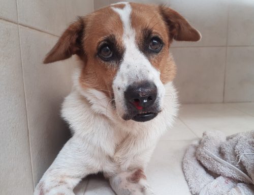
Signs That Need Immediate Evaluation
In young pets, eye problems can progress rapidly. Same-day evaluation is appropriate for:
- Any sudden cloudiness or color change in the eye
- One eye held shut or squinting repeatedly
- Thick, colored, or excessive discharge
- Visible swelling, bulging, or protrusion of the eye
- Asymmetry between the two eyes
- Pawing persistently at the face or rubbing the eye on furniture
- Pupils that are unequal in size or unresponsive to light changes
Appointment requests for urgent presentations are accommodated as quickly as possible.
Supporting Healthy Eye Development at Home
While specialist evaluation is necessary for clinical concerns, daily home habits support healthy eye development:
- Keep play environments free of sharp objects and materials that could penetrate the eye
- Monitor interactions with other young animals, particularly rough play around the head
- Maintain good nutrition with a complete and balanced diet through the growth period
- Keep the periocular area clean of discharge to prevent skin irritation and infection
- Schedule consistent wellness visits through the first year so your primary care veterinarian can monitor development
Any change from your pet’s established baseline around the eyes, including new discharge, altered appearance, or behavioral changes suggesting vision difficulty, is a reason to reach out rather than wait.
Frequently Asked Questions
When should I have my puppy or kitten’s eyes examined by a specialist?
Your primary care veterinarian will refer when they identify findings that warrant specialist evaluation. Some breeders request a CAER (Companion Animal Eye Registry) exam at 7 to 8 weeks for breed-specific screening. Any concern about eye appearance, development, or vision at any age warrants a call.
What is a CAER exam?
A Companion Animal Eye Registry examination is a standardized ophthalmic exam performed by a board-certified veterinary ophthalmologist to screen for hereditary eye conditions. Results can be registered with OFA. CAER examinations are part of what we offer to breeders and families wanting hereditary screening for their pets.
Can congenital eye conditions be treated?
It depends on the condition. Some require surgery, some are managed medically, some are monitored, and some have no effective intervention but benefit from assessment to define prognosis. The goal of early evaluation is understanding what is present and what it means for the individual animal.
Clear Vision Starts Early
The window of visual development in puppies and kittens is short, and conditions that are manageable when caught early can produce permanent damage when missed. Our team at Veterinary Vision Center brings the diagnostic precision of board-certified ophthalmology to every evaluation.
Request an appointment or contact us at (318) 797-5522 to schedule a pediatric eye evaluation.
Leave A Comment