
We don’t get much snow here in Shreveport, Louisiana, but you may still have a snow globe as part of your winter decorations. What if you were gazing at your snowglobe one day, and noticed that the serene cabin surrounded by snow-laden fir trees has been joined by a garish, fluorescent-lit, 24-hour convenience store. This change to your snow globe is analogous to a tumor that suddenly pops up in the snow globe of your pet’s eye, and is unsightly, disruptive, and problematic.
Intraocular tumor look-alikes in pets
Although most eye masses are cancerous, some processes can cause changes to the eye structure’s appearance, or look like a mass, but may or may not be a concerning cancerous tumor. Dr. Pierce can help differentiate intraocular tumors from:
- Iris nevi — Also known as iris freckles or iris melanosis, these dark, pigmented spots that appear on your pet’s iris cause no problems. However, a spot that grows, becomes thickened, or causes inflammation may actually be an iris melanoma or melanocytoma, which are discussed later.
- Uveal cysts — These round, sometimes free-floating structures are typically somewhat translucent and vary in color from dark brown or black to clear. They look odd, but pose little risk to your pet.
Intraocular tumors in dogs
Primary (i.e., arising from the eye itself) and secondary (i.e., spread to the eye from a distant location) intraocular tumors can be found in dogs, most commonly:
- Melanocytoma and melanoma — These tumors of the eye’s pigment-producing cells may be benign (i.e., melanocytoma) in 85% to 90% of cases, or malignant (i.e., melanoma) in 10% to 15% of cases. The most common dog intraocular tumors, they are dark brown to black, raised or nodular in appearance, and can be seen on the iris’ surface, or pushing the iris forward.
- Ciliary body adenomas and adenocarcinomas — These tan to pink masses, which can be seen protruding through the pupil or pushing the iris forward, originate from the ciliary body, a structure close behind the iris that produces the aqueous humor (i.e., the fluid in the front part of the eye). They seem more common in Labrador retrievers and golden retrievers, and can be benign (i.e., adenoma) in 85% of cases, or malignant (i.e., adenocarcinoma) in 15% of cases.
- Metastatic tumors — These secondary tumors occur because of the spread of lymphoma, histiocytic sarcoma, mammary adenocarcinoma, hemangiosarcoma, malignant melanoma, or osteosarcoma from other body parts, and make up 14.8% of all intraocular tumors in dogs. Dogs who have systemic signs or tumors in both eyes are more likely to have a metastatic tumor than a primary intraocular tumor.
Intraocular tumors in cats
Cats can also suffer from primary or secondary intraocular tumors, but their tumors are more likely malignant than in dogs. Some common cat intraocular tumors are:
- Melanoma — Cats may start out with a harmless color change to the iris (i.e., iris nevi) that may later spread, thicken, and become malignant. Melanoma has a 55% to 66% metastasis rate and is the most common feline intraocular tumor.
- Post-traumatic sarcoma — Blunt or penetrating lens trauma or ongoing eye inflammation (i.e., uveitis) can cause the lens cells to become malignant and form an aggressive, highly invasive tumor that can sometimes travel up the optic nerve to the brain. Because of this tumor risk, cats with known ocular trauma or uveitis should have regular eye exams by an ophthalmologist, and blind or significantly damaged eyes should be removed to prevent tumor formation.
- Metastatic tumors — Lymphoma is the most common cause for secondary intraocular tumors, especially in cats who are infected with feline leukemia virus (FELV) or feline immunodeficiency virus (FIV), although the other tumors listed for dogs can also occur.
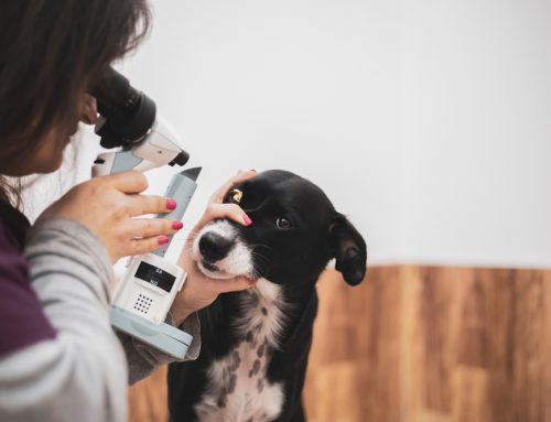
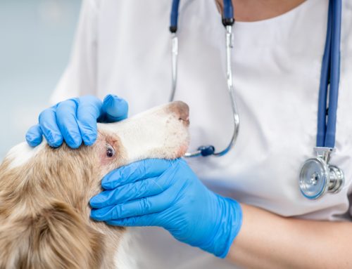
Diagnosis of intraocular tumors in pets
Not all affected pets have an immediately visible tumor, so some are initially evaluated because they have glaucoma (i.e., high eye pressure), hyphema (i.e., bleeding in the eye), uveitis, or eye pain, and the diagnostic work-up for those conditions reveals a tumor. Dr. Pierce will always perform a complete ophthalmic exam to evaluate the front of the eye and the retina, measure eye pressure, and use specialized testing such as an eye ultrasound, CT, or MRI to find and further evaluate the tumor’s size, location, and effects. He can make an educated guess about tumor type, or in some cases, insert a small needle into an anesthetized pet’s eye to obtain a sample of tumor cells, but the most definitive diagnosis comes from removing the tumor-containing eye and submitting it for a biopsy.
Treatment of intraocular tumors in pets

The tumor type and its effect on your pet will help guide treatment. Treatment for pets who are experiencing glaucoma, uveitis, or eye pain that cannot be well controlled, or pets suspected to have aggressive or malignant tumors, is typically eye removal. The eye is then submitted for biopsy to guide decisions about additional therapy such as chemotherapy or radiation. Visual dogs with melanocytomas that do not appear to be causing inflammation or pain can possibly be monitored, or sometimes their tumor can be destroyed using a special laser. Secondary intraocular tumors can be addressed as part of the treatment for the primary cancer type, and may or may not necessitate eye removal.
At Veterinary Vision Center, we aren’t in the business of snowglobe renovations, but we are experts at diagnosing and managing your pet’s intraocular tumors. Give us a call to put the snowglobe of your pet’s eye in Dr. Pierce’s capable hands.


I found a lot of useful information on your site. Thank you for the valuable information. Author of the blog: https://espiar-gratis.com/