When you bring your dog in for a red, inflamed eye, tick-borne disease isn’t usually at the top of your mental checklist. But Lyme disease, Ehrlichia, Rocky Mountain Spotted Fever, and Anaplasma can all produce ocular signs that range from uveitis to retinal hemorrhage to sudden blindness, and missing the systemic connection means treating the eye while the underlying infection continues unchecked. In regions where ticks are active, including across much of Louisiana and the wider Southeast, the systemic diseases they carry are always in the differential for unexplained ocular inflammation.
Veterinary Vision Center in Shreveport is a board-certified ophthalmology referral practice, and our work at the intersection of systemic medicine and ocular disease means we see the full picture when tick-borne disease affects the eye. Dr. Pierce and our team are experienced in identifying the ocular manifestations of infectious disease and coordinating with general practitioners to address both the eye and the underlying cause. Our services include advanced ophthalmic diagnostics for complex cases. Request an appointment to discuss a referral.
What Matters Most
- Tick-borne diseases including Ehrlichia, Rocky Mountain Spotted Fever, Lyme, and Anaplasma can all cause eye inflammation, retinal bleeding, sudden blindness, or other ocular changes, and the eyes are sometimes the first place the disease becomes visible.
- Up to 37% of dogs with ehrlichiosis developed ocular signs in one study, which is why a red or cloudy eye in a tick-exposed dog deserves a workup that looks beyond the eye itself.
- Effective treatment requires addressing both the systemic infection (usually doxycycline) and the ocular inflammation at the same time; treating one without the other rarely produces a good outcome.
- Tick prevention and prompt removal of attached ticks are the most reliable way to protect your pet’s vision from these diseases, since outcomes are best when infection is prevented or caught early.
How Do Tick-Borne Diseases Cause Eye Problems in Pets?
Lyme disease and the eyes is one example of a broader pattern: tick-borne infections affect the eye through several distinct mechanisms.
- Direct invasion of ocular tissue occurs when the infectious organism reaches eye structures and produces local damage. The eye is highly vascular, and bloodborne pathogens reach ocular tissue readily.
- Immune complex deposition triggers inflammation inside the eye. The immune system’s response to the systemic infection produces antibody-antigen complexes that deposit in the uveal tract, driving uveitis even when the organism itself isn’t present in the eye.
- Vasculitis disrupts blood supply to the delicate structures of the eye. The retinal blood vessels in particular are vulnerable to inflammatory damage from tick-borne diseases.
- Systemic hypertension from kidney disease (a complication of several tick-borne infections) can damage the retina through pressure-related vascular changes.
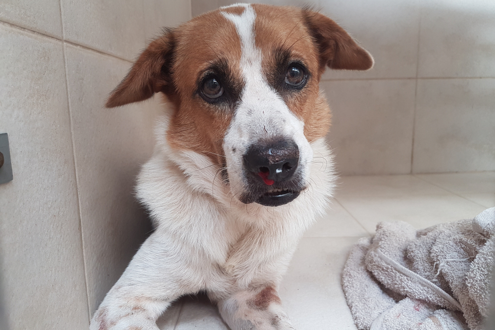
The ocular pain associated with intraocular inflammation often presents as squinting, photophobia and eye pain, and reluctance to be in bright light. Pets with uveitis are uncomfortable, and the discomfort often gets attributed to other causes before the underlying inflammation is recognized.
What Are the Common Eye Symptoms of Tick-Borne Disease?
Tick-borne disease can produce a recognizable cluster of ocular findings. The presentations overlap across the different organisms, but knowing what each one looks like helps make sense of why a tick panel might be on the workup list for a dog with new eye symptoms.
- Uveitis: inflammation inside the eye, often causing redness, squinting, and a cloudy appearance. Managing uveitis requires both controlling the inflammation and identifying the underlying cause, since untreated uveitis can progress to secondary glaucoma, retinal detachment, cataract formation, and permanent vision loss.
- Retinal detachment: separation of the retina from underlying tissue, often producing sudden blindness. Detachment can develop quickly in severe cases and requires prompt intervention if vision preservation is possible.
- Conjunctivitis and chemosis: redness and swelling of the tissues lining the eyelids. Often the most visible early sign, easy to mistake for an isolated eye problem when it is actually one piece of a systemic picture.
- Optic neuritis: inflammation of the optic nerve, which can disrupt the transmission of visual information to the brain and produce sudden vision changes even when the rest of the eye looks relatively normal.
- Hyphema and hemorrhage: bleeding directly into the chambers of the eye, often appearing as visible blood in the front of the eye or red-tinged discoloration. Common when tick-borne disease has caused low platelet counts.
- Corneal edema and opacities: swelling or cloudiness of the surface of the eye, sometimes visible as a hazy, blue-grey appearance over the front of the eye.
Any one of these findings warrants a workup. Several appearing together raise suspicion for a systemic cause and shape both the diagnostic plan and the urgency of treatment.
Which Tick-Borne Diseases Cause Which Eye Problems?
Different tick-borne pathogens favor different patterns of ocular involvement. The patterns are not absolute (a dog with ehrlichiosis can present with any of the findings above) but knowing what each disease typically does helps make sense of an eye exam in the context of a tick panel.
Ehrlichiosis (Tick Fever)
Ehrlichia is one of the tick-borne diseases most likely to cause significant ocular complications. Up to 37% of dogs with ehrlichiosis develop ocular signs, and the eye changes are sometimes the first noticeable indicator of the disease before lameness, lethargy, or fever bring families in.
The ocular pattern in ehrlichiosis frequently includes:
- Uveitis, often the leading sign
- Retinal disease including hemorrhage and vasculitis
- Severe bleeding within the eye driven by the thrombocytopenia (low platelet counts) that ehrlichiosis produces
- Chronic or recurring uveitis in some dogs that persists after the acute infection has been treated, suggesting an ongoing immune-mediated process
A red or cloudy eye in a dog with tick exposure is reason enough to run the full panel, even when the dog seems systemically well.
Rocky Mountain Spotted Fever (RMSF)
Rocky Mountain spotted fever occurs across much of North America despite the regional name, including Louisiana and the Southeast. The disease drives vasculitis throughout the body, and the eye, with its dense network of small vessels in the retina and uvea, is particularly vulnerable.
RMSF is associated with:
- Eye swelling and conjunctivitis, often among the first visible signs
- Uveitis with the standard signs of intraocular inflammation
- Hyphema (bleeding inside the eye)
- Retinal edema and retinal hemorrhage
- Sudden vision loss when severe vasculitis disrupts the structural integrity of the retina or causes detachment
Ocular disease from RMSF can develop rapidly. The window for effective treatment is narrow; pets with significant ocular involvement need immediate evaluation and treatment to preserve vision.
Lyme Disease
Lyme disease is the most widely recognized tick-borne illness in North America, transmitted by the black-legged tick. While most attention focuses on the joint pain and lameness commonly associated with Lyme, the ocular manifestations matter.
Lyme less commonly affects the eyes than some of the other tick-borne diseases, but when it does, the patterns include:
- Uveitis, the most common ocular sign
- Conjunctivitis with redness and tearing
- Retinal detachment in more severe cases
- Posterior segment changes including retinitis, retinal hemorrhage, and optic neuritis
Ocular signs from Lyme disease can appear during active infection or in the weeks following treatment. New eye symptoms in your dog who has recently been treated for Lyme disease warrant prompt re-evaluation; the signs can indicate ongoing immune-mediated inflammation that requires its own management even when the bacterial infection has been cleared.
Anaplasmosis
Anaplasmosis typically produces less severe ocular involvement than ehrlichiosis but can cause steroid-responsive uveitis. The mechanism is similar to ehrlichiosis (both organisms infect blood cells and can produce thrombocytopenia), and co-infection with both organisms is common. Tick-borne disease panels test for multiple organisms simultaneously because ticks frequently transmit more than one pathogen at a time.
For complex cases requiring coordinated specialty care, our services support general practitioners managing the systemic component while ophthalmology addresses the ocular manifestations.
How Do You Recognize Eye Symptoms That May Signal Tick-Borne Disease?
The clinical signs that warrant a same-day call to the clinic:
- Eye redness in one or both eyes
- Squinting or holding the eye partially closed
- Excessive tearing or unusual discharge
- Cloudiness or haze within or on the surface of the eye
- Visible blood inside the eye or on the conjunctiva
- Pupil size or symmetry changes (one pupil larger or smaller than the other, pupils not responding to light)
- Light sensitivity or avoidance of bright environments
- Behavioral signs of reduced vision including bumping into things, hesitancy on stairs, or unwillingness to navigate unfamiliar areas
These signs can look similar across several different conditions. Distinguishing tick-borne disease as the underlying cause from other reasons for ocular emergencies requires examination rather than home observation.
Acute glaucoma, corneal ulcers, traumatic injury, and immune-mediated diseases not related to ticks can all produce overlapping presentations. Prompt evaluation determines which category applies and shapes the urgency of treatment.
The Diagnostic Workup for Tick-Borne Ocular Disease
Diagnosing the cause of uveitis or other ocular signs in your pet with suspected tick-borne disease requires both an ophthalmic evaluation and a systemic workup.
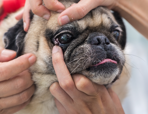
The ophthalmic examination includes:
- Tonometry to measure intraocular pressure (low pressure suggests uveitis; high pressure can indicate secondary glaucoma)
- Slit-lamp biomicroscopy of the anterior segment to evaluate the cornea, anterior chamber, iris, and lens
- Anterior chamber assessment for inflammatory cells or blood
- Pupillary light reflex testing to evaluate retinal and optic nerve function
- Dilated fundic examination to evaluate the retina, optic nerve, and vitreous
- Fluorescein staining to identify any concurrent corneal ulceration
The systemic workup includes:
- Complete blood count evaluating red cells, white cells, and platelet counts (low platelets suggest tick-borne disease specifically)
- Chemistry panel assessing organ function and screening for kidney involvement
- Urinalysis evaluating kidney function and protein loss
- Tick-borne disease panel testing for multiple organisms simultaneously
- Blood pressure measurement, since hypertension can drive retinal damage independent of direct ocular infection
How Is Ocular Disease From Tick-Borne Infections Treated?
Effective treatment requires addressing both the systemic infection and the ocular inflammation at the same time. Treating only the eye leaves the underlying infection in place; treating only the infection allows the eye damage to continue unchecked.
The antibiotic component is doxycycline as the foundation for most tick-borne bacterial infections. Doxycycline penetrates well into ocular tissue, reaches effective concentrations against the relevant pathogens, and is generally well-tolerated. Treatment durations typically extend 21 to 28 days for acute presentations and longer for chronic cases.
The ocular treatment approach addresses inflammation directly. Treatment of intraocular inflammation typically involves:
- Topical anti-inflammatories to reduce inflammation in the anterior segment
- Cycloplegic agents that dilate the pupil and reduce iris-to-lens adhesions during active inflammation
- Topical corticosteroids when immune-mediated inflammation is the primary driver and the corneal surface is intact
- Systemic corticosteroids in selected cases (introduced cautiously when active infection is present to avoid suppressing the immune response needed to clear the infection)
- Anti-glaucoma medications if intraocular pressure rises during treatment
Giving your pet eye medications is its own learning curve, especially when multiple drops are needed throughout the day. We provide instruction during your visit and are available to answer questions during the treatment course.
Complications to Watch for During and After Treatment
Several ocular complications can develop during active uveitis or after treatment without proper monitoring:
- Secondary glaucoma develops when inflammation interferes with normal aqueous outflow, raising intraocular pressure to damaging levels. Glaucoma can develop quickly and requires prompt adjustment of the treatment plan.
- Cataract formation can result from chronic uveitis as inflammation damages the lens. Some uveitis-induced cataracts are amenable to surgical management; others affect overall visual function more substantially.
- Iris-to-lens adhesions (synechiae) develop when inflamed iris tissue sticks to the lens during active inflammation. These adhesions can be permanent and may interfere with pupil function long after the active inflammation resolves.
- Retinal detachment can occur in severe cases, particularly with significant retinal vasculitis or hemorrhage. Detachment requires prompt intervention if vision preservation is to be possible.
- Permanent vision loss is the worst-case outcome of severe or neglected disease. The risk is real but largely preventable with prompt diagnosis and aggressive coordinated treatment.
Monitoring intraocular pressure throughout treatment is essential. Recheck appointments aren’t optional; they’re how we catch developing complications early.
Tick Prevention as the Best Protection for Eye Health
Preventing tick-borne disease is the most reliable way to avoid tick-borne ocular complications. Consistent year-round tick prevention with veterinarian-recommended products provides the foundation. Year-round prevention is appropriate in most regions including Louisiana, where tick activity extends across most of the year.
Additional risk-reduction strategies:
- Thorough tick checks after outdoor activity, which identify attached ticks before they’ve had time to transmit disease (the transmission window varies by pathogen and prompt removal substantially reduces infection risk)
- Prompt and correct removal of attached ticks using fine-tipped tweezers, grasping the tick close to the skin, and pulling straight upward without twisting
- Awareness of high-risk habitats including wooded areas, tall grass, and brush
- Annual screening through tick-borne disease testing to identify subclinical infections before they progress
Discussion with your primary veterinarian about the most appropriate preventive product for your pet’s lifestyle and regional exposure risk supports informed product selection.

Frequently Asked Questions About Tick-Borne Eye Disease in Pets
My dog tested positive for Lyme but has no symptoms. Should I worry about the eyes?
Subclinical Lyme infections occasionally progress to symptomatic disease including eye involvement. Routine eye examinations during follow-up help catch any developing ocular involvement early.
How quickly should I act if my pet’s eye looks abnormal?
Same-day evaluation is appropriate for any new significant eye change. Eye conditions can progress rapidly, and the difference between same-day treatment and waiting a few days can be the difference between vision preservation and permanent loss.
Can my pet recover full vision after tick-borne uveitis?
Many pets achieve excellent vision recovery with prompt and aggressive treatment. Outcomes depend on how quickly treatment was initiated, the severity of the inflammation, and whether complications developed.
Will my pet need to stay on eye medications long-term?
Treatment duration varies by case. Acute uveitis typically requires several weeks of intensive therapy with gradual tapering as the eye improves. Some pets with chronic inflammation require lower-dose maintenance therapy indefinitely.
Do cats get tick-borne ocular disease too?
Cats develop tick-borne disease less commonly than dogs but are not exempt. Toxoplasmosis is a more common cause of ocular inflammation in cats than tick-borne disease specifically, but the diagnostic and treatment principles overlap substantially.
Protecting Your Pet’s Vision From Tick-Borne Disease
Pets with a history of tick-borne illness should have any new eye symptoms evaluated promptly rather than monitored at home. Combined systemic and ophthalmic treatment gives most pets an excellent chance of preserving vision and comfort, but the timeline matters; the window for the best outcomes is narrower than most people assume.
If your pet has a history of tick-borne disease and is showing eye changes, or if you’d like to discuss tick-borne disease screening in the context of unexplained ocular signs, request an appointment at Veterinary Vision Center. Our team is ready to evaluate, diagnose, and coordinate treatment with your primary veterinarian.


Leave A Comment