Tear Staining, Sun Sensitivity, and Ocular Care in Light-Coated Pets
White-faced pets are undeniably adorable, but come with health risks. Families may start noticing things like reddish-brown staining beneath the eyes, pink skin around the eyelids that looks a little raw after a sunny afternoon, or eyes that seem smaller or different in structure than other dogs in the household. These observations are worth taking seriously, because reduced pigmentation around the eyes does change how those tissues respond to light, UV exposure, and certain inherited conditions.
The encouraging part is that most eye conditions affecting white and light-coated pets are manageable when identified early. Veterinary Vision Center, led by board-certified ophthalmologist Dr. Pierce, is located inside University Veterinary Hospital in Shreveport, LA. Our services include advanced diagnostics, surgical therapies, and comprehensive management for everything from hereditary eye defects to UV-related corneal disease, delivered with a family-style approach. Contact us at (318) 797-5522 to schedule an ophthalmology consultation.
Why Do White and Light-Pigmented Pets Get Tear Stains?
What Causes Tear Stains, and When Should You Call the Vet?
Tear staining is one of the first things you may notice with a white or light-coated pet, and it is one of the most common reasons families start looking into ocular health for the first time. The brownish-red discoloration beneath the eyes is caused by porphyrins, pigment compounds naturally present in tears. Any dog or cat can develop tear stains; they’re just much more noticeable on white pets. When tears overflow onto pale fur and remain wet against the skin, porphyrins oxidize and stain. The staining itself is a cosmetic issue, but the reason the tears are overflowing in the first place is the more important question.
- Tear stains are most prominent in breeds with shallow eye sockets, prominent eyes, or heavy facial hair, including Maltese, Bichons, Poodles, Shih Tzus, and brachycephalic breeds like Bulldogs and Pugs. The anatomy of these faces shortens the normal drainage path for tears and creates surfaces where moisture collects.
- Eyelids that don’t close properly or rub on the eye and hair around the eyes that touches or wicks against the cornea adds to the problem, simultaneously causing irritation that stimulates more tearing and creating a channel that holds moisture against the skin.
- Blocked or narrow tear ducts, eye lashes touching the eye, allergies, dental disease affecting the roots near the eye, and corneal irritation can all drive excessive tearing.
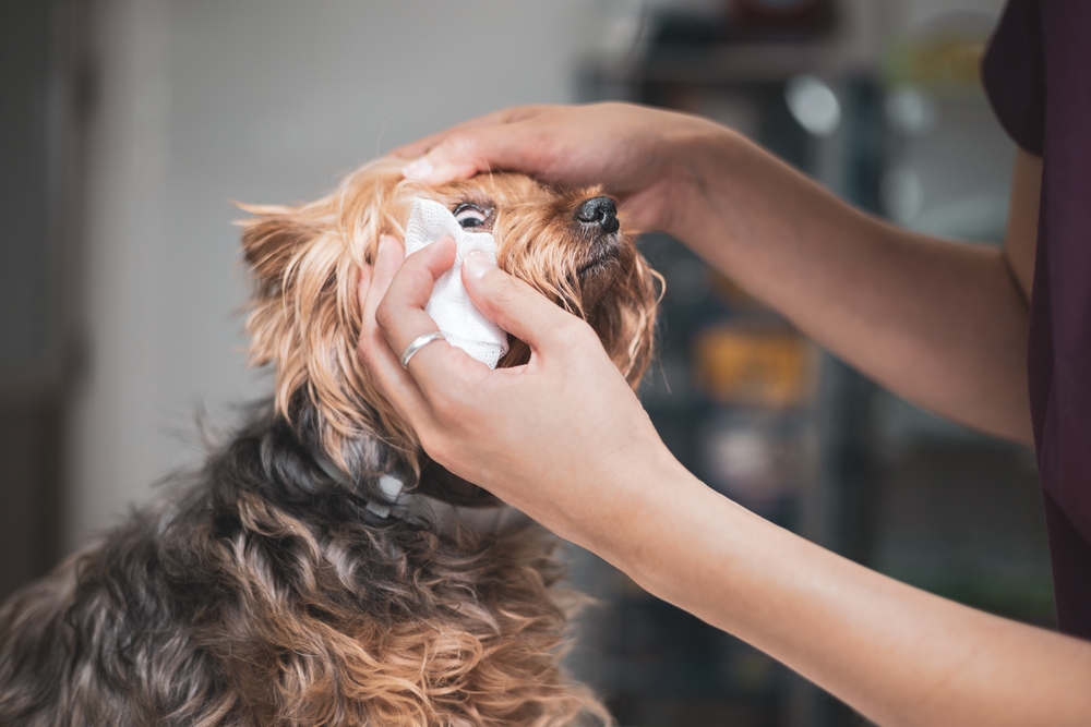
In light-coated pets, secondary yeast and bacterial growth in the moist fur adds odor and can cause skin irritation beneath the stain. Consistent daily cleaning of the face with a veterinarian-approved wipe or saline-moistened cloth keeps the area drier and reduces secondary infections. Trimming the hair around the eye area so it does not contact the cornea is equally useful.
The signs that move tear staining from a cosmetic concern to a medical one include a change in discharge color from clear to yellow or green, visible redness or cloudiness of the eye, squinting, increased rubbing at the face, or any sudden change in the pattern or amount of tearing. When those signs appear, evaluation is needed promptly. Our team assesses the full drainage system, corneal surface, and eye health during ophthalmic exams to identify whether staining has a treatable underlying cause.
What Genetic Eye Conditions Affect Merle and White-Coated Breeds?
Merle Gene Eye Defects: What You Should Know
The merle gene creates the dappled, patchy coat patterns found in Australian Shepherds, Great Danes, Collies, and Dachshunds. Dogs that inherit a single copy generally develop normally. The serious problems arise with double merle dogs, those who inherit two copies, one from each parent, because double merle status is strongly associated with congenital eye defects.
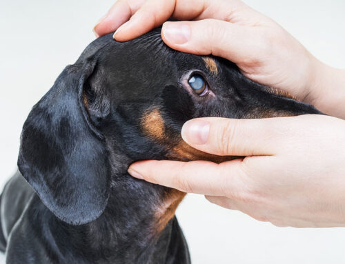
Double merle dogs are often predominantly or entirely white, and merle eye defects in these animals can include microphthalmia (abnormally small eyes), iris colobomas (gaps or holes in the iris structure), lens displacement, retinal abnormalities, and varying degrees of vision impairment up to complete blindness. The condition results from disruption to the melanocyte migration that is essential for normal eye development. The affected structures may look visibly different, with smaller eyes, unusual iris coloring, or a slightly sunken appearance, or the defects may be internal and only detectable through examination.
Responsible breeding practice requires that two merle-pattern dogs are never bred together, because every puppy from that pairing has a 25 percent chance of inheriting the double merle genotype. Genetic testing is available and appropriate for any breeder working with affected breeds. If you have adopted or purchased a double merle dog, early ophthalmology evaluation establishes exactly what your pet can and cannot see and guides appropriate management.
What Is Microphthalmia, and Can These Pets Still Live Well?
Microphthalmia describes a condition in which one or both eyes are abnormally small, and it is frequently accompanied by other developmental abnormalities of the eye structures. Microphthalmia and ocular dysgenesis represent a spectrum of conditions where the eye did not develop normally in utero, and affected pets may have associated cataracts, corneal abnormalities, lens displacement, or retinal dysplasia alongside the reduced eye size.
Breeds where this condition appears more frequently include Australian Shepherds, Collies, Doberman Pinschers, and Miniature Schnauzers. The connection to excessive white coat patterning and merle genetics means that double merle dogs are disproportionately represented among affected patients.
You may notice some of these signs:
- Recessed eye position
- Prominent third eyelid elevation
- Irregular or off-center pupil
- Reduced responses to visual stimuli.
The vision these pets have at birth is generally the vision they will have, as the structural defects are congenital rather than progressive. Most pets with microphthalmia adapt remarkably well to whatever level of vision they possess, relying on scent, hearing, and spatial memory in ways that allow them to navigate familiar environments confidently. The goal of veterinary care is to understand the full extent of the defects, manage any associated conditions like corneal ulcers or cataracts that might be treatable, and equip you with realistic expectations and practical home management strategies. We provide exactly that level of individualized assessment and guidance through our ocular condition evaluations.

Does Sun Exposure Affect Pets With Light Pigmentation?
UV Risk and Sunburn in White and Pale-Skinned Pets
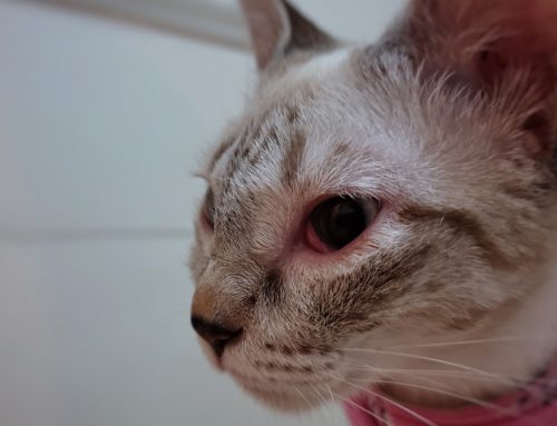
Pigmentation in skin and the tissues around the eye serves a protective function against ultraviolet radiation. Pets with white coats and pink skin around the eyelids, nose, and ears lack that natural barrier, which makes them genuinely more susceptible to UV damage than their more pigmented counterparts. In Louisiana, where UV intensity is high for most of the year, this is a practical and ongoing consideration for affected patients.
Sunburn in pets most commonly appears on the ear tips, nose bridge, and the skin around the eyes in light-pigmented animals. Acute sunburn produces redness and tenderness. Chronic sun exposure over months and years can lead to actinic keratosis, a thickening and crusting of damaged skin, and in some cases squamous cell carcinoma, a type of skin cancer that is locally invasive and requires surgical management. White cats, white Boxers, Bull Terriers, and Dalmatians are among the most consistently affected patients, though any pet with pink periocular skin should be considered at elevated risk.
Practical UV protection for light-pigmented pets includes limiting unshaded outdoor time during peak UV hours (roughly 10 a.m. to 4 p.m.), providing covered resting areas outdoors, and applying pet-safe sunscreen to the ear tips, nose, and skin around the eyes when sun exposure is unavoidable. Products formulated specifically for pets avoid the ingredients, including zinc oxide and para-aminobenzoic acid, that are toxic when licked. Signs of sun damage that warrant veterinary attention include redness or scaling that does not resolve, crusting lesions on the ear tips or nose, and any tissue that appears thickened, discolored, or ulcerated.
The eyes themselves are also vulnerable. Solar-induced keratitis, a progressive inflammation and pigmentation of the cornea in response to UV exposure, is seen in pets with inadequate periocular pigment. We evaluate corneal health as part of every ophthalmic examination and can identify early UV-related changes before they progress.
Can Protective Eyewear Actually Help?
Protective eyewear designed specifically for dogs provides a practical option for pets with significant UV sensitivity, corneal conditions, or those who regularly spend extended time in high-exposure environments like boats, hiking trails, and snow. Quality canine goggles block UV radiation, shield the corneal surface from debris and wind, and in pets with corneal disease or prior surgical procedures, reduce environmental irritation during outdoor activity.
Not every dog takes to wearing goggles without resistance, but gradual introduction using positive reinforcement makes acceptance achievable for many patients. Starting with short indoor sessions, using high-value treats, and building duration slowly tends to work far better than forcing the process. The fit matters: properly fitted eyewear stays in place without pressing on the eye surface or slipping during activity. For pets who cannot tolerate goggles, the most effective alternatives are strategic outdoor timing to avoid peak UV hours and consistent use of shaded spaces.
What Does Routine Eye Care Look Like for At-Risk Breeds?
How Often Should Light-Pigmented Pets Have Eye Exams?
For breeds with known hereditary ocular conditions, a baseline ophthalmology examination early in life, ideally before one year of age, establishes what is normal for that individual and identifies any congenital defects before they cause secondary complications. Annual follow-up examinations are appropriate for most at-risk breeds, with more frequent monitoring recommended for pets with active conditions or progressive diseases like hereditary cataracts or progressive retinal atrophy.
A comprehensive ophthalmic examination at Veterinary Vision Center includes Schirmer tear testing to measure tear production, tonometry to measure intraocular pressure, corneal evaluation, assessment of the lens for clarity, fundic examination of the retina and optic nerve, and where indicated, advanced imaging or electroretinography. This level of detail identifies problems at stages where intervention is most effective.
Monitoring at Home: What to Watch Between Appointments
Daily observation is genuinely useful. Knowing what your pet’s eyes look like normally makes you the first to notice when something changes, and early reporting consistently improves outcomes. Watch for cloudiness or a bluish haze to the eye, redness of the white of the eye or eyelid lining, squinting or holding one eye partially closed, changes in discharge, and behavioral signs of vision difficulty like hesitation at doorways or bumping into furniture in unfamiliar spaces.
Keeping the facial area clean, trimming hair that contacts the eye, and documenting changes with photos to share with us all contribute to effective at-home monitoring. When any warning signs appear, prompt contact is the right response.
We believe that quality of life is at the center of everything we do, and that includes equipping you with practical tools for supporting your pet through vision changes. Request an appointment to discuss your light-pigmented pet’s specific needs, or schedule a consultation to establish a baseline for breeds at elevated ocular risk.
Eye Health Is Worth Protecting: Starting Now
White and light-pigmented pets bring a particular kind of beauty into a home, and they deserve the attentive, individualized eye care that matches it. Whether the concern is tear staining that never fully clears, a breed with known hereditary risks, sun sensitivity in a dog who loves afternoons outside, or a newly adopted double merle whose vision you want to understand better, we provide the specialist expertise to evaluate and manage it.
Dr. Pierce and our team approach every patient with the same standard: gold-standard care delivered like family, because that is what every pet deserves. Call (318) 797-5522 or contact us to schedule an evaluation.


Leave A Comment