When pets develop eye problems, treatment often falls into two categories: medical management or surgery. The choice depends on the condition, the pet, and what you as the pet owner can manage at home. What works for one pet or family may not work for another. Choosing the right path starts with understanding what each option can and cannot do for your pet’s specific condition, and what makes sense for your situation.
At Veterinary Vision Center in Shreveport, LA, we provide gold-standard ophthalmology care in a comfortable, family-style setting. Our board certified eye doctor guides you through options, from medical management to advanced surgery, so you can make decisions with confidence. If you are curious about the treatment options available to you for your pet’s eye condition, reach out. We’re here to help you understand and make choices that make sense for your pet.
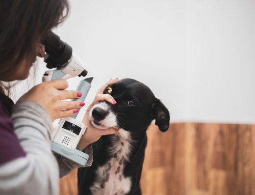
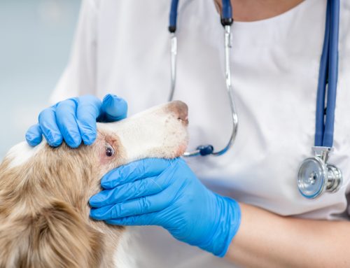
How Does a Veterinary Ophthalmologist Evaluate Eye Problems?
A thorough exam is the most important first step. We start with a hands-on assessment and simple, pet-friendly tests, using advanced tools when needed. A careful physical examination of the eye checks the structures that protect sight and comfort.
We assess the eyelids, conjunctiva, and surrounding tissues to spot irritation, masses, or structural issues. Corneal staining reveals ulcers or scratches. Measuring eye pressure screens for glaucoma, a painful condition that can steal vision quickly. We check pupil response and perform a dilated exam to evaluate the retina and optic nerve. When changes are not easily seen, we turn to imaging and specialty equipment.
Corneal Ulcers: Scratches and Sores on the Eye’s Surface
Corneal ulcers are open sores on the clear front surface of the eye. They range from minor scratches to deep, vision-threatening wounds. Causes include trauma, foreign material, dry eye, and eyelid abnormalities that allow lashes to rub the surface.
When medication is enough: Many superficial ulcers heal well with antibiotic drops, pain relief, lubrication, and an e-collar to prevent rubbing. Most simple ulcers improve within days to a week with consistent treatment and monitoring.
When surgery is recommended: Deep or complicated ulcers that extend into the deeper layers of the cornea, or ulcers that risk perforating the eye, often need surgical support. Procedures like grid keratotomy help non-healing superficial ulcers by creating small scratches that encourage the surface to adhere and heal. For deeper wounds, a conjunctival graft brings blood supply directly to the damaged area to stabilise the cornea and promote repair. You can read more about what to expect from corneal surgery at our hospital, or visit our page on corneal ulcers for additional detail. If your pet is squinting or has a scratch, book an eye exam so we can determine the depth and plan accordingly.
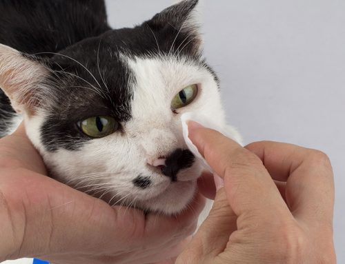
Dry Eye: When Tear Production Falls Short
Dry eye (keratoconjunctivitis sicca) means the eye is not making enough tears to protect the surface. Without adequate moisture, the cornea becomes inflamed, painful, and vulnerable to ulcers and infection.
When medication is enough: Most dogs with dry eye respond well to daily tear stimulants (like cyclosporine or tacrolimus) combined with artificial tears and lubricants. Medication is typically lifelong, but most pets settle into a manageable routine. Regular rechecks fine-tune dosing and catch changes early. Our team helps you build a schedule that works for your household, and tips for administering eye medications make the process smoother.
When surgery is recommended: In cases where medication cannot maintain adequate tear production and the eyes remain chronically irritated, a procedure called parotid duct transposition may be considered. This surgery redirects a salivary duct to deliver moisture to the eye surface. It is not common, but for pets who do not respond to medical therapy, it can be a valuable option.
Eyelid Abnormalities: Structural Problems That Irritate the Eye
Structural eyelid issues cause chronic rubbing, poor eye protection, or constant irritation that drops alone cannot fix. These conditions are common in certain breeds and tend to worsen over time if left uncorrected.
Entropion causes the eyelid to roll inward so lashes scrape the cornea with every blink. Cherry eye is a prolapsed tear gland that appears as a pink mass in the corner of the eye and often needs repositioning to preserve tear production. Eyelash disorders like distichiasis or ectopic cilia create ongoing surface irritation.
When medication is enough: Lubricating drops and antibiotics can manage mild irritation or protect the eye while waiting for a planned surgical correction. In very young animals, temporary tacking sutures may be used while the face is still growing.
When surgery is recommended: Most structural eyelid problems ultimately need surgical correction because the anatomy driving the irritation does not resolve on its own. Our guide to veterinary eyelid surgery walks through eyelid procedures in plain terms, including what recovery looks like and how we protect comfort throughout healing.
Eyelid and Eye Tumors: Growths That Need Attention
Growths on or around the eye are relatively common, especially in older pets. Eyelid tumours in dogs are often benign but can irritate the cornea as they grow, while cats tend to develop more aggressive eyelid masses that need prompt attention. Tumours can also develop inside the eye itself, including melanomas, lymphoma, and other cancers and tumours of the eye.
When monitoring or medication is enough: Very small eyelid growths that are not touching the cornea can sometimes be monitored with regular rechecks. Anti-inflammatory drops may help manage secondary irritation while we track changes.
When surgery is recommended: Most eyelid tumours that are growing, rubbing the eye surface, or suspected to be aggressive benefit from early surgical removal. Smaller tumours are simpler to remove with better cosmetic and functional results, which is why we encourage evaluation sooner rather than later. You can learn more about when eyelid tumour surgery is necessary and what the process involves on our site.
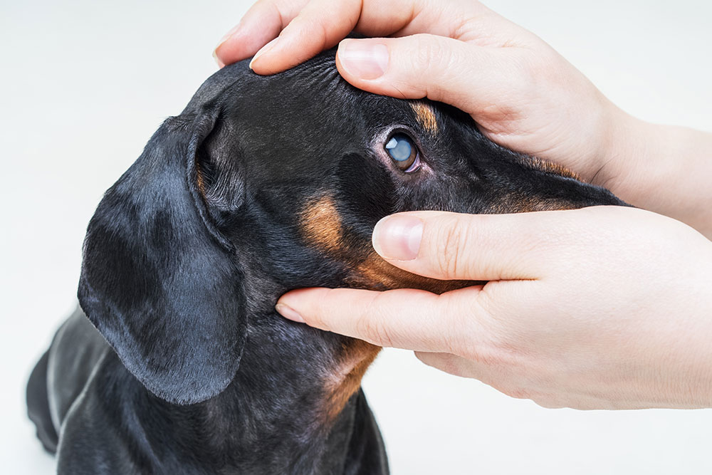
Cataracts: Cloudy Lenses That Block Vision
A cataract is a clouding of the lens inside the eye that blocks light from reaching the retina. Cataracts can develop from diabetes, genetics, aging, inflammation, or trauma. They range from small opacities that barely affect vision to dense clouds that cause functional blindness.
When medication is enough: There are no drops that dissolve cataracts. However, anti-inflammatory eye drops are used to manage the inflammation that cataracts cause inside the eye (called lens-induced uveitis), and these drops may be recommended whether or not surgery is pursued. Medical management helps keep the eye comfortable and reduces the risk of secondary complications like glaucoma.
When surgery is recommended: When a cataract significantly impairs vision and the eye is otherwise healthy, surgery is the only way to restore sight. The procedure involves breaking up and removing the cloudy lens, often replacing it with an artificial one. Early to mid-stage cataracts generally have better surgical outcomes than advanced ones. Pre-surgery testing confirms the retina and optic nerve are functioning well. You can read about what to expect before, during, and after cataract surgery to understand the full process.
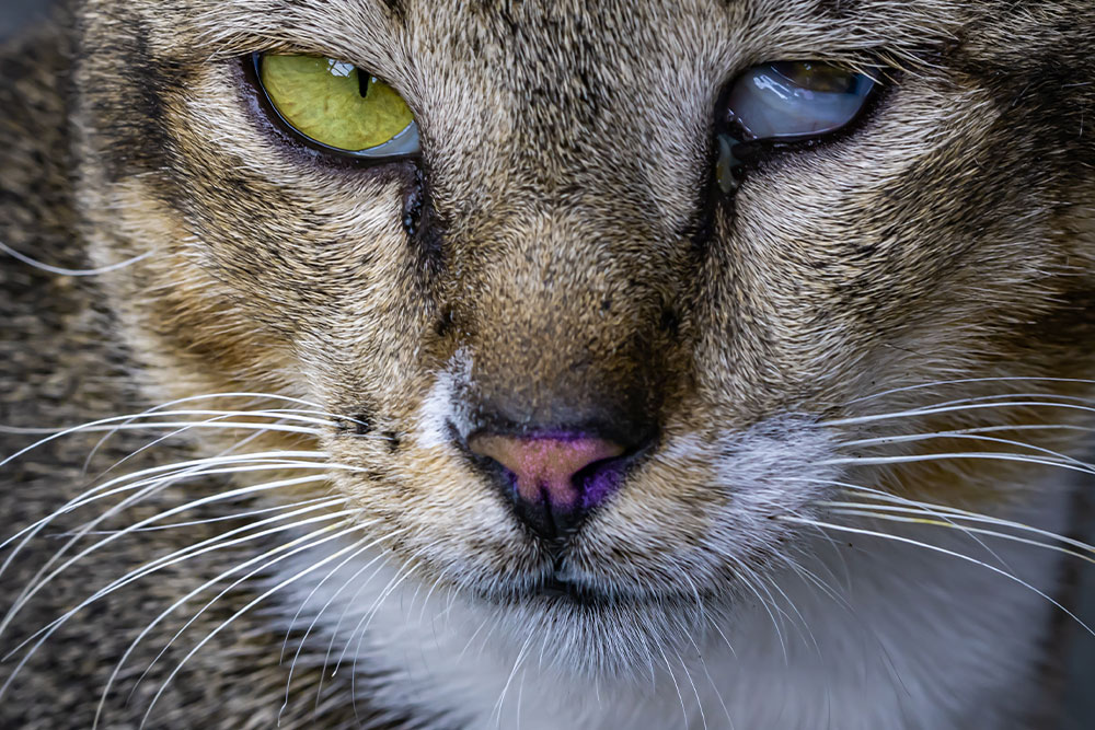
Glaucoma: Dangerous Pressure Inside the Eye
Glaucoma is elevated pressure inside the eye that damages the optic nerve and retina. It is painful and can permanently destroy vision quickly if not controlled.
When medication is enough: Many cases begin with pressure-lowering eye drops and sometimes oral medications. When pressure responds well and vision is intact, ongoing medical management with close monitoring can maintain comfort. We track pressure regularly and adjust medications as the disease evolves.
When surgery is recommended: Glaucoma is progressive, and many cases eventually require surgical intervention to protect remaining vision or relieve pain. Vision-sparing procedures may help earlier in the disease, while comfort-focused surgeries (including laser procedures or implants to reduce fluid production) are considered when vision has been lost but pain persists. Our page on what to expect from glaucoma surgery outlines the options and recovery process.
Lens Luxation: When the Lens Shifts Out of Place
When the lens moves from its normal position, it can block fluid drainage and trigger sudden, painful glaucoma. Many cases of lens dislocation need urgent treatment. Some terrier breeds are genetically predisposed.
When medication is enough: If the lens shifts backward (posterior luxation) and pressure remains normal, medical monitoring with pressure-lowering drops may be sufficient in the short term.
When surgery is recommended: Anterior luxation (lens moving forward) is an emergency. Surgery to remove the displaced lens reduces pain and prevents secondary damage. If your pet shows acute eye pain, a suddenly larger-looking eye, or rapid cloudiness, contact us immediately.
Retinal Detachment and Conditions Affecting the Back of the Eye
The retina is the light-sensitive tissue lining the back of the eye. Retinal detachment occurs when the retina separates from the underlying tissue, causing partial or complete vision loss. High blood pressure, immune-mediated disease, infection, and trauma are common causes.
When medication is enough: When retinal detachment is caused by high blood pressure or inflammation, treating the underlying condition with blood pressure medications or anti-inflammatory drugs can sometimes allow the retina to reattach. Early intervention gives the best chance of preserving vision.
When surgery is recommended: Surgical reattachment is possible in some cases but depends heavily on the cause, duration, and extent of the detachment. Not all detachments are surgical candidates, which is why prompt evaluation matters.
Uveal Cysts: Usually Harmless, Occasionally a Problem
Uveal cysts are fluid-filled structures that float inside the eye. Most are incidental findings that do not affect vision or comfort.
When monitoring is enough: The majority of uveal cysts require nothing more than periodic observation during routine eye exams.
When intervention is recommended: If a cyst grows large enough to block the pupil or affect vision, deflation or removal can be considered. Decisions are based on visual impact and comfort.
Exophthalmos, Proptosis, and Bulging Eyes
Exophthalmos is the forward displacement of the eye within the socket, giving it a bulging appearance. Causes include infection or abscess behind the eye, tumours in the orbital space, and inflammation. It is different from breed-related prominent eyes seen in brachycephalic dogs.
Treatment depends entirely on the underlying cause. Infections may respond to antibiotics and drainage, while tumours often require advanced imaging and surgical planning. Because the eye and surrounding structures are under pressure, prompt evaluation protects both vision and comfort.
Proptosis occurs when the eye actually comes out of the socket beyond the eyelids. It can occur from trauma, and it’s more common in flat-faced pets with large, bulging eyes. It is considered an emergency and if caught soon enough, the eye can be replaced and the eyelids temporarily surgically closed to protect the eye while it heals. If not, removal of the eye may be needed. For pets with especially flat faces and bulging eyes, a medial canthoplasty can reshape the inner corner of the eye opening, providing better coverage of the eye and making it less likely proptose.
When Is Removing the Eye the Right Decision?
Enucleation (surgical removal of the eye) is recommended when an eye is blind and painful, or when a tumour or severe trauma makes saving the eye unsafe. It is never a first choice, but for pets living with chronic, unmanageable pain, it is often the most compassionate option. Most pets adjust remarkably well and return to normal activity quickly.
What Factors Shape the Treatment Recommendation?
Every pet is different. We consider overall health and anaesthesia safety by reviewing history, running screening tests, and balancing risks with benefits. Conditions like heart or kidney disease may shift the plan toward medical management. Quality of life and comfort guide every decision, and these conversations are always transparent and centred on your pet’s wellbeing.
Owner lifestyle matters too. Many eye conditions require attentive home care: multiple daily drops, an e-collar, and frequent rechecks. For pets that need eye drops more frequently than their owner can provide, or for pets who are extremely stressed when receiving eye drops and are facing long-term treatment needs, surgery may be a better option. For others, it may make more sense to stick with long-term medical management. We’ll guide you through the short-term and long-term outlooks and choices for your pet’s diagnosis. Our guide to ophthalmic surgery aftercare outlines what to expect with surgery so you can decide what is feasible before committing to a plan.
Frequently Asked Questions
What signs mean my pet needs an eye exam? Redness, squinting, cloudiness, pawing at the eye, discharge, sudden clumsiness, or light sensitivity.
How fast should I act if I see sudden changes? Immediately. Sudden pain, bulging, rapid cloudiness, or vision loss is an emergency. Contact us right away.
Can cataracts be treated with drops? No. Drops manage inflammation caused by cataracts, but surgery is the only way to restore vision.
Will my pet need lifelong drops for dry eye? Usually, yes- and multiple times a day. Most pets do well with daily medications and periodic rechecks to adjust dosing.
How do pets adjust after losing an eye? Most dogs and cats adapt quickly and return to normal activity. Pets rely heavily on their other senses, and quality of life is typically excellent.
Protecting Your Pet’s Vision Through Informed Choices
Both medical management and surgery have important roles in protecting sight and comfort. The right choice depends on the condition, its severity, your pet’s overall health, and what works for your family. Acting early almost always improves the outcome.
If you have questions or want guidance tailored to your pet, request an appointment or contact us. Our team is here to help your pet see the world comfortably.

Leave A Comment