When Eyelid Surgery Requires a Specialist: Complex Entropion, Ectropion, and Concurrent Eye Conditions
If your pet’s regular veterinarian referred them to a veterinary ophthalmologist for eyelid surgery, it is worth understanding what that referral actually means. It does not mean something has gone wrong or that the situation is alarming. It means that eyelid surgery, while it looks straightforward on the surface, involves a level of surgical precision, anatomical judgment, and concurrent condition management that is reliably better handled by someone who does it exclusively. The margin for error is genuinely small: a millimeter difference in the amount of tissue removed determines whether a pet is overcorrected, undercorrected, or corrected. Get it wrong in either direction and you are planning a second surgery.
At Veterinary Vision Center, our board-certified veterinary ophthalmologist evaluates and treats the full spectrum of eyelid conditions, including cases that have already been tried elsewhere, cases with multiple problems compounding each other, and cases where the lid position looks like the whole problem but is actually part of a larger picture. Request an appointment or contact us to schedule a consultation.
Why Do General Practitioners Refer Eyelid Cases?
Most primary care veterinarians are very comfortable evaluating entropion and ectropion and initiating early management. What prompts a referral is usually one or more of the following:
- The eyelid anatomy is complicated by heavy skin folds, facial compression, or asymmetry
- The pet has already had a tacking procedure or a prior surgical attempt that did not fully resolve the problem
- There is significant corneal damage that needs to be assessed and managed alongside the lid correction
- The pet has concurrent eyelid conditions such as distichiasis or trichiasis that complicate the surgical picture
- The condition involves both entropion and ectropion in the same eye
- The pet’s age, breed, or overall health status makes the anesthetic and surgical approach less straightforward
- The eyelid position has improved but the eye is still showing signs of pain or surface damage
These are not rare exceptions. They are the cases we see regularly, and evaluating them requires both specialized imaging and the experience to interpret what the findings mean for surgical planning.
The Complexity of Concurrent Eyelid and Ocular Surface Conditions
One of the most common reasons eyelid surgery does not produce the expected improvement is that the lid position was not the only problem, or not the primary problem, to begin with.
Distichiasis and Trichiasis: When the Lashes Are the Issue
Eyelash problems frequently coexist with entropion and ectropion. Distichiasis refers to lashes that emerge from abnormal follicles along the lid margin, often pointing directly at the cornea. Trichiasis describes normally positioned lashes that are misdirected inward, usually due to surrounding skin folds rather than the lid margin itself rolling. Both cause the same kind of corneal friction as entropion, and both can be missed on a standard examination if the lid margin is not evaluated under magnification.
The significance for surgical planning: removing skin to correct lid roll does not address a misdirected lash. If distichiasis or trichiasis is part of the picture, those problems need to be identified and addressed separately, often in the same surgical session, or the pet will continue to experience corneal irritation after the lid position is corrected.
Dry Eye (KCS) as a Complicating Factor
Keratoconjunctivitis sicca (KCS), commonly called dry eye, is a condition where the lacrimal glands produce insufficient tear volume. It is particularly common in breeds also prone to eyelid abnormalities, including Bulldogs, Cocker Spaniels, and West Highland White Terriers.
A pet with significant KCS and entropion who undergoes lid correction may still have a chronically unhealthy corneal surface because the tear film is inadequate, not because the lid position is wrong. If KCS is missed preoperatively and the pet continues to struggle after lid correction, the natural assumption is that the surgery did not work when in reality the tear film problem was never addressed. The ocular tests performed at a specialist consultation, including Schirmer tear testing, are specifically designed to catch this before surgery rather than after.
Corneal Ulcers and Surface Damage at the Time of Referral
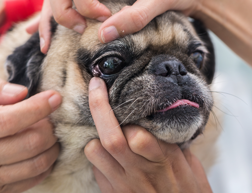
Many pets arrive at our practice with active corneal damage. Corneal ulcers develop when persistent lid friction breaks down the corneal epithelium, ranging from superficial surface abrasions to deep stromal ulcers approaching the inner layers of the eye. The presence and depth of ulceration directly affects the surgical timeline.
A superficial ulcer may be manageable alongside lid correction. A deep or infected ulcer may need to be stabilized first, with lid surgery delayed until the cornea is safe to operate around. Some pets arrive with corneal scarring from months or years of unaddressed friction, which requires honest discussion about how much vision impact has occurred and whether any of it is reversible. The lid surgery addresses the cause; it does not reverse established scar tissue.
Diamond Eye: Entropion and Ectropion in the Same Lid
Some breeds, particularly those with very loose facial skin and heavy folds, develop a combination of entropion at the outer and inner corners and ectropion in the middle of the lower lid. This creates the characteristic appearance that gives the condition its name. Saint Bernards, Basset Hounds, and Clumber Spaniels are classic examples.
Correcting diamond eye is not a matter of simply addressing one component. Tightening the outer lid without accounting for the middle portion, or vice versa, creates a new imbalance. The surgical plan has to account for the full arc of lid position across its length, which requires precise pre-surgical mapping and calibration. The eyelid disorders that occur in combination are consistently harder to correct than isolated entropion or ectropion in a breed without significant facial redundancy.
When Prior Surgery Did Not Fully Resolve the Problem
Revision cases are a meaningful portion of our surgical caseload. Pets arrive having had a previous entropion correction where the lid improved but did not fully resolve, or where improvement was seen initially and the lid rolled inward again as healing progressed.
The reasons vary. In young animals, facial structure continues to change after the first correction, and what was accurate at six months may need adjustment as the face matures. In dogs with heavy skin folds, remaining redundant tissue can continue to pull on the corrected area. In some cases, the initial correction underestimated the extent of the entropion, which is easier to do than it sounds: topical anesthetic applied to the eye relaxes the lid and can make the degree of rolling appear less severe than it is under normal conditions.
If your pet has already had a correction and is still symptomatic, we will evaluate what was done, where the lid currently sits, and what the cornea shows before recommending a path forward.
Breed-Specific Surgical Complexity
The same procedure is performed very differently in a Shar-Pei with extreme skin redundancy than in a Labrador Retriever with a straightforward lateral entropion. Different breeds have different needs.
Brachycephalic breeds, including Boxers, Bulldogs, and French Bulldogs, frequently develop medial canthal entropion, where the rolling occurs at the inner corner of the eye rather than along the full lid margin. This location is particularly challenging because the inner canthus lies close to the lacrimal puncta, the small drainage openings that move tears into the nasal passages, and surgical technique must account for that anatomy to avoid creating a secondary drainage problem.
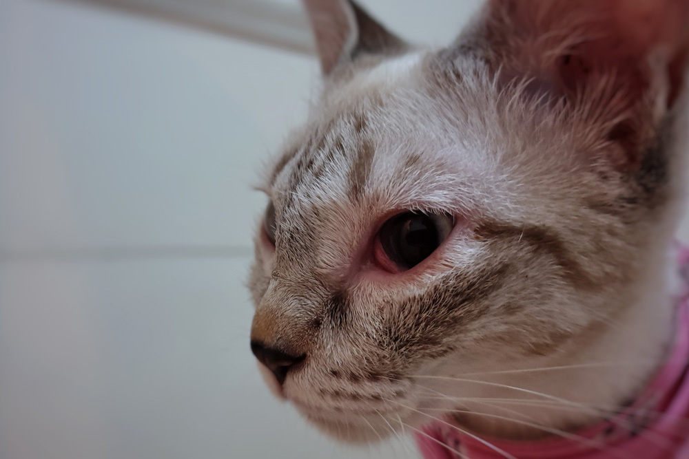
Entropion in cats typically develops differently than in dogs. Rather than a hereditary structural abnormality, feline entropion often develops as a secondary consequence of chronic squinting from another cause, such as herpesvirus-associated ocular surface disease, chronic conjunctivitis, or corneal disease. In these cats, correcting the lid position without treating the underlying ocular surface disease that caused the squinting leads to recurrence. Flat-faced breeds including Persians and Himalayans can also develop primary structural entropion, particularly at the medial canthus, but even in these patients a thorough evaluation of the overall ocular surface is necessary before a surgical plan is finalized.
The Specialist Diagnostic Process
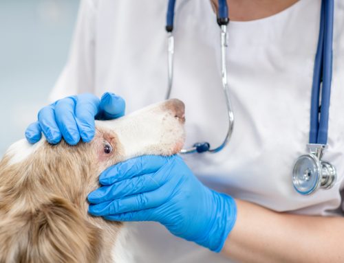
When your pet is seen at Veterinary Vision Center, the evaluation is built specifically around identifying everything that is contributing to the problem, not just the most obvious component.
The examination includes ocular tests that build a complete picture before any surgical decisions are made:
- Lid position assessment before and after topical anesthetic, to distinguish true structural entropion from spastic entropion driven by pain and squinting
- Schirmer tear test to document tear production and identify KCS as a concurrent factor
- Fluorescein staining to map corneal ulcers, erosions, and surface damage invisible to the naked eye
- Magnified slit-lamp examination of the lid margins, lash emergence points, corneal surface, and conjunctival tissue
- Evaluation of lid tension and skin redundancy across the full arc of the eyelid to plan tissue removal accurately
For pets with existing corneal ulcers, the depth and characteristics of the ulcer are assessed to determine whether lid surgery can proceed immediately or whether corneal management needs to come first. For revision cases, the prior surgical result is carefully documented before planning what approach will address what remains.
Surgical Approaches for Complex Cases
The eyelid surgery used to correct entropion and ectropion is not a single procedure applied uniformly. The approach is specific to the location, degree, and contributing anatomy of each case.
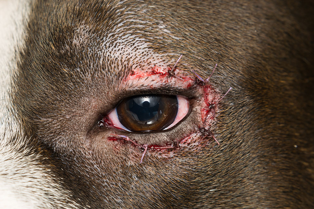
For straightforward lateral entropion, a Hotz-Celsus procedure removes a precisely measured ellipse of skin below the lid margin. For medial canthal entropion, the technique is modified to preserve the drainage anatomy at the inner corner. For diamond eye, the correction has to address both the rolled and the sagging components of the lid in sequence. For cases with concurrent distichiasis, electroepilation or cryotherapy to the affected follicles is performed alongside the lid correction.
In young animals still growing into their faces, temporary eyelid tacking can protect the cornea and give the lid margin a chance to settle before permanent surgery. Some puppies correct adequately with tacking alone. Others require permanent correction once growth is complete. Tacking is not a substitute for surgical correction in adult animals, and it is not appropriate in cases where corneal damage is already significant.
What Signs Indicate Your Pet Needs an Evaluation
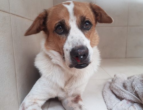
Signs of eye pain in pets are not always obvious. Reluctance to have the face touched, avoidance of bright areas, intermittent squinting, or crankiness near the head can all indicate significant ocular discomfort. More visible signs include:
- Persistent tearing or a watery eye that has not resolved with treatment
- Pawing at the face or rubbing along furniture or carpet
- Cloudiness, haziness, or visible color change on the corneal surface
- Thick, colored, or recurring eye discharge
- A visibly rolled-in lid margin or a sagging lower lid
- Any pet that has already had eyelid surgery and is still showing signs of irritation
Administering eye medications can manage symptoms temporarily, but medication does not correct the structural problem generating them. Chronic reliance on eye drops when surgery is what is needed allows secondary corneal damage to accumulate in the background.
Recovery in Complex Cases
In uncomplicated cases, swelling peaks in the first 24 to 48 hours, pets show relief from friction symptoms within the first day or two, and sutures come out at the 10 to 14 day recheck. The full correction settles by four to six weeks.
Complex cases may follow a different course. Pets with concurrent corneal ulcers require additional recheck appointments to confirm the corneal surface is healing. Pets treated for distichiasis alongside their lid correction may need follow-up assessment of lash regrowth. Revision cases require careful documentation of the final lid position at multiple points during healing.
The e-collar is non-negotiable regardless of case complexity. The most common reason surgical sites are disrupted is the e-collar being removed briefly to “give the pet a break.” It stays on continuously until the recheck. If you have questions about recovery, you can always reach out to our team.
Frequently Asked Questions
My pet had entropion surgery but is still squinting. What is happening?
This is one of the most common reasons pets are referred to us after a prior procedure. The usual explanations are that the correction was incomplete, a concurrent condition like KCS or distichiasis was not identified and treated, or there is residual corneal damage that needs separate management. A specialist evaluation identifies which combination applies.
Can entropion recur after surgical correction?
In breeds with significant facial redundancy or ongoing facial development, it can. This is why staged corrections are sometimes recommended in young dogs, and why revision surgery exists as an option when initial correction is incomplete. Permanent correction is the outcome in most cases.
Why does the eyelid need to be evaluated before and after anesthetic drops?
Applying topical anesthetic relaxes the lid and reduces spasm. In some patients, what looks like moderate entropion before anesthetic resolves almost entirely afterward, indicating that a significant spastic component is present. This matters because removing tissue to correct spasm alone can produce overcorrection once the underlying cause of the squinting is treated. The pre- and post-anesthetic lid assessment distinguishes structural entropion from spastic entropion, which changes the surgical plan.
My cat’s entropion keeps coming back after being treated. Why?
In cats, recurrent entropion frequently indicates that the underlying cause of the squinting has not been fully addressed. Feline herpesvirus, chronic conjunctivitis, and other ocular surface diseases cause persistent squinting, and persistent squinting causes the lid to roll. Correcting the lid without managing the ocular surface disease that is driving the squinting typically leads to recurrence.
Getting the Right Evaluation the First Time
Eyelid conditions are among the most correctable problems in veterinary ophthalmology, and the outcomes in well-selected, carefully executed cases are consistently good. The cases that do not go as planned are almost always the ones where concurrent conditions were not identified before surgery, where the correction was not calibrated precisely to the individual anatomy, or where the underlying cause of a secondary entropion was not addressed.
If your pet has been referred for eyelid evaluation, or if they have already had a correction that did not fully resolve the problem, we want to see the full picture before making recommendations. Request an appointment at Veterinary Vision Center in Shreveport, inside University Veterinary Hospital, or give us a call. We’re here to help.
Leave A Comment