
Quincy Quarter Horse and Polly Pocket Pony are munching on hay in adjoining barn stalls.
Quincy: Hey, Polly Pocket, did I tell you that I finally got someone to tell my owner that I can have all the carrots I want?
Polly Pocket: Wait, what? How did that happen? I want unlimited carrots, too!
Quincy: Well, I’m still a little puzzled by how it all went down, but here’s the scoop. Things seemed to be looking a little fuzzy, and my owner noticed that my eye was cloudy, so she called Dr. Pierce. He visited me yesterday, looked at my eye, and diagnosed me with something called immune mediated carrot-itis. I don’t really know what some of the big words meant, but I do know what carrots are, so I think carrot-itis means that I have a carrot deficiency. Dr. Pierce told my owner to treat my carrot-itis daily, so obviously that means she should bring me carrots every day. I’m not really sure how eating carrots has anything to do with my blurry vision, but I’m not going to ask too many questions. Look! There is my owner now. I hope she remembered that I prefer organic baby carrots.
Poor Quincy is going to be disappointed to find out that he actually has immune mediated keratitis (IMMK), which has nothing to do with carrots. However, he does need his eye medicated daily, so perhaps he can talk his owner into giving him a carrot, peppermint, or other treat at the same time.
Definition of equine IMMK
Keratitis refers to cornea inflammation, and the designation “immune mediated” indicates that this inflammation occurs when the horse’s immune system mounts an abnormal response to a foreign protein, cornea component, or infectious agent. IMMK occurs in one eye in 85% of cases, but can occur in both eyes, especially with the eosinophilic keratitis type. IMMK tends to occur in horses older than 12, with no known sex or breed predisposition.
Types of IMMK in horses
IMMK can be classified into five types:
- Epithelial IMMK — This is the least common form and the most superficial. Affected horses may have a rough corneal appearance or scattered small opaque dots on the cornea.
- Superficial stromal IMMK — This is the most common IMMK type, comprising 45% of all cases, and is characterized by blood vessels branching across the cornea surface and a yellow-to-white corneal opacity.
- Mid-stromal IMMK — This type makes up 27% of cases and typically has straighter vessel growth, with a more opaque cornea compared with superficial stromal IMMK.
- Endothelial IMMK — This chronic, slowly progressive form makes up 23% of IMMK cases. Affected horses have a blue appearance to some or all of the cornea, and may occasionally have bubbles on the cornea surface that rupture to form corneal ulcers.
- Eosinophilic keratitis — This condition is sometimes classified as IMMK but does have some differences compared with other IMMK types. The condition occurs in early or late summer, sometimes affects multiple horses in a barn, and may involve a white plaque on the cornea surface and/or irritation of the conjunctiva with a thick white discharge. Eosinophilic keratitis often spontaneously resolves in 8 to 12 weeks, unlike other IMMK types, which do not typically go away on their own. Eosinophilic keratitis is also driven by eosinophils, a different cell type than seen in other IMMK forms.
Signs a horse may have IMMK
With the exception of eosinophilic keratitis, which may cause moderate discomfort, most horses with IMMK do not seem to experience much, if any, eye pain or discomfort. The eye’s appearance will vary depending on the type, but most cases have some degree of corneal opacity, which may wax and wane or progress over time. IMMK typically hinders the horse’s vision to some extent as the disease progresses.
IMMK diagnosis in horses
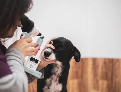
Dr. Pierce will perform a complete ophthalmic exam before diagnosing your horse with IMMK. He will use a handheld microscope called a slit lamp to evaluate the eye’s surface and front chamber contents, and may stain the eye to look for ulcers, measure the eye pressure to screen for glaucoma, and visually evaluate the retina. To help distinguish IMMK from a corneal ulcer or infection, he may also examine the cornea cells or perform a culture. This is important because, with the exception of corneal ulcers being painful and IMMK being nonpainful, the eye appearance can be similar in all diseases, but IMMK treatment can make a corneal ulcer or infection much worse.
Treatment for horses with IMMK

The exact protocol will depend on the IMMK type and eye appearance, but generally treatment involves topical anti-inflammatory or immune-modulating (i.e., altering the immune response) drugs. In some cases, Dr. Pierce may perform a surgery called a superficial keratectomy to remove the diseased cornea portion. Researchers at North Carolina State College of Veterinary Medicine are investigating the use of stem cell therapy to treat IMMK, with some promising results so far, so this may become a viable treatment option.
Prognosis for horses with IMMK
Prognosis varies, depending on the IMMK type and therapy response. Some horses maintain acceptable vision, while others, especially those with endothelial IMMK, may lose significant vision. The earlier the IMMK diagnosis, the sooner treatment can be started in hopes of maintaining as much vision as possible, so promptly contact Veterinary Vision Center if you or your family veterinarian suspect your horse has IMMK.
Quincy may not get carrots every day, but he should still be grateful that Dr. Pierce diagnosed his IMMK, and started him on the correct medications to maintain his sight as long as possible, so his doting owner can enjoy many more rides. If your horse is experiencing eye problems, Veterinary Vision Center is the place to call.

Leave A Comment