
Cloudy Eyes in Pets: What Corneal Dystrophy Means for Vision
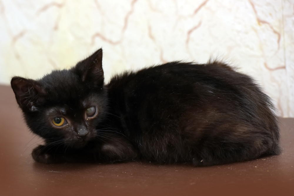
The moment you notice a faint cloudiness or haze developing in your pet’s eyes, a wave of concern washes over you. Are in pain? Can still see you clearly? Without a thorough ophthalmic exam, it’s impossible to know.
This common change could be a sign of corneal dystrophy, a genetic disorder causing abnormal deposits in the transparent layer of the eye. Though often painless, this condition can progress and potentially affect your pet’s sight if not monitored closely. Understanding the early signs is the first step a pet owner can take to work with a veterinarian on a plan to preserve eye health and prevent future complications.
At Veterinary Vision Center, we see firsthand how small changes in a pet’s eyes can have a big emotional impact on their families. As a specialty practice dedicated entirely to ophthalmology, we provide comprehensive evaluations, advanced diagnostics, and surgical options for dogs, cats, and exotic pets. Our mission is simple: to care for every pet as if they were our own- and to protect the special bond between you and your companion.
Understanding Corneal Dystrophy in Pets
Corneal dystrophy refers to an inherited condition that affects the cornea, the clear outer layer of the eye. It develops when lipids, minerals, or proteins accumulate within specific corneal layers, causing the surface to appear cloudy or opaque. This buildup is typically non-inflammatory and occurs in both eyes, though one may appear worse than the other.
Corneal dystrophy often begins as a faint haze near the center of the cornea, progressing slowly over time. Many pets show no pain or vision loss, but in some cases, the deposits can scatter light enough to interfere with visual clarity. Early evaluation through specialized eye exams and imaging helps confirm the diagnosis and track changes safely over time.
How Hereditary Eye Diseases Develop
Genetics play a significant role in how corneal dystrophy and related conditions form. Certain breeds- including Siberian Huskies, Boston Terriers, and Cocker Spaniels- are more prone to hereditary eye disease due to inherited structural or metabolic differences. These predispositions influence when symptoms appear and how quickly the condition may progress.
Understanding canine eye health allows veterinarians to tailor screening schedules for at-risk breeds and detect subtle changes before they affect vision. At Veterinary Vision Center, we use advanced slit-lamp biomicroscopy and optical imaging to monitor your pet’s eyes with precision, adjusting care as needed to preserve comfort and sight.
Recognizing the Types and Symptoms of Corneal Dystrophy
Corneal dystrophy is categorized based on which corneal layer is affected- each type presenting slightly differently.
Epithelial and Stromal Corneal Changes
Epithelial dystrophy affects the cornea’s surface layer. It may appear as a soft haze or faint white spots and rarely causes discomfort. Stromal dystrophy, on the other hand, occurs deeper in the cornea and can produce a more noticeable opacity or shimmer. These forms are usually cosmetic but should be monitored regularly to ensure they do not progress.
Regular ophthalmic evaluations at our specialty clinic allow early detection and tracking, giving owners peace of mind through professional monitoring and clear guidance.
Endothelial Corneal Changes
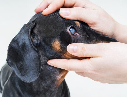
When the innermost layer of the cornea is affected, the condition is known as corneal endothelial degeneration. The endothelium regulates corneal hydration; when it weakens, fluid can accumulate within the cornea, leading to swelling, bluish discoloration, tearing, or discomfort. Without treatment, this can impair vision or predispose pets to ulcers.
Our board-certified ophthalmologist uses advanced diagnostics like specular microscopy and pachymetry to monitor these changes and determine the best approach for preserving comfort and vision.
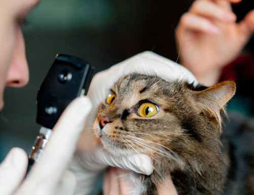
How Veterinarians Diagnose Corneal Dystrophy
Diagnosis involves a combination of clinical expertise and specialized technology. Using a slit-lamp biomicroscope, our team can evaluate each layer of the cornea and distinguish dystrophy from other diseases. Fluorescein staining ensures there are no ulcers or scratches on the surface, while genetic background and breed history offer important context.
This comprehensive evaluation is critical for accurate differentiation between dystrophy, degeneration, and inflammatory disorders. Our in-house imaging capabilities and expert care allow for precise monitoring and treatment planning- all within one compassionate, family-style environment.
Treatment and Management Options
While many pets with corneal dystrophy require only monitoring, certain cases may benefit from medical or surgical management. Our approach focuses on comfort, eye protection, and preventing secondary complications.
Therapies may include:
- Lubricating drops or ointments to maintain moisture and reduce irritation
- Antioxidant or anti-inflammatory support if mild discomfort occurs
- Routine rechecks to track changes over time
- Surgical intervention for severe endothelial involvement or recurring ulceration
If topical medications are prescribed, our team provides hands-on guidance on how to apply eye drops correctly and safely at home.
At Veterinary Vision Center, every treatment plan is customized to the pet’s individual condition and comfort needs. From initial diagnosis to long-term management, our focus is always on improving your pet’s quality of life.
Differentiating Corneal Dystrophy from Other Eye Conditions
Because many eye issues cause cloudiness, it’s important to distinguish hereditary dystrophy from inflammatory or infectious causes. Conditions like conjunctivitis often include redness, discharge, or squinting, while corneal ulcers cause significant pain and tearing- but both can cause cloudiness similar in appearance to corneal dystrophy.
If your pet suddenly develops eye discomfort, redness, or visible discharge, same-day evaluation is essential. Our veterinary corneal ulcer care provides specialized diagnostics and treatment for these urgent issues, ensuring fast relief and expert attention.
When to Seek Immediate Help
Managing hereditary conditions requires consistency and partnership. We recommend annual or semiannual exams depending on the severity and breed. Each visit includes updated imaging, photos, and a review of visual behavior. These records help us fine-tune your pet’s care plan and maintain clarity and comfort throughout their life.
Although corneal dystrophy typically progresses slowly, any change in cloudiness, behavior, or comfort warrants attention. Signs like rubbing at the eye, squinting, increased tearing, or sudden opacity may indicate complications.
Our team provides prompt evaluations and imaging to determine whether intervention is needed. The earlier these changes are addressed, the better the long-term outcome for vision and comfort.
Protecting Your Pet’s Vision with Proactive Care
Corneal dystrophy doesn’t have to stand in the way of a happy, active life. With compassionate care and routine follow-ups, most pets live comfortably without major complications. The key is staying proactive- monitoring for subtle changes and seeking expert guidance early.
If you notice any cloudiness, irritation, or behavioral changes in your pet’s eyes, schedule an appointment today or contact our team. At Veterinary Vision Center, we’re here to provide world-class eye care in a family-style setting- because your pet’s vision, comfort, and happiness mean the world to us.


Leave A Comment