
X-rays and ultrasound are common veterinary medicine tools. Imaging tests allow us to see inside a pet’s body in a non-invasive manner and get the answers we need to help them heal. Modern imaging has advanced, and we now use imaging techniques that specifically look inside and around the eyes. The Veterinary Vision Center team routinely uses eye imaging tests to diagnose disease and evaluate overall eye health. Here is an overview of how and why our team recommends ocular imaging.
Diagnosing eye problems in pets
Dogs and cats can potentially develop many different eye diseases and injuries that can cause long-term discomfort or vision loss. Diagnosing and treating eye problems early can help reverse or prevent eye damage, or at least slow down the progression of a particular disease. A general practice veterinarian can diagnose some eye problems, but many require complex examination techniques and diagnostics for a more complete picture.
Referral to a veterinary ophthalmologist is the next step for a pet who has unusual eye issues or isn’t responding to standard treatments. Ophthalmologists use an array of techniques to arrive at a diagnosis, including:
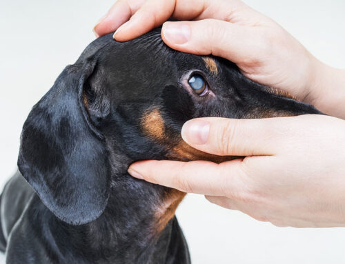
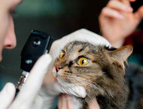
- Ophthalmic exam — A handheld microscope allows the ophthalmologist to examine the structures on the outside and front inside portions of the eye in great detail. A handheld lens and light source shone through the pupil allow a brief exam of the retina, which lines the inside back of the eye.
- Big three diagnostics — These diagnostics are performed during the exam and include tonometry to check eye pressure, a Schirmer tear test to determine tear production, and fluorescein dye to check for eye surface scratches.
- Specialized tests — If more information is needed, ophthalmologists can perform additional in-office tests, such as electroretinography to check retinal function or gonioscopy to examine the internal eye fluid drain (i.e., the iridocorneal angle).
- Imaging tests — If other tests don’t provide enough information, imaging tests can help the ophthalmologist see inside the eye in greater detail and from additional angles. Common eye imaging tests include ultrasound, computed tomography (CT), and magnetic resonance imaging (MRI).
Benefits of ocular imaging in pets
Ocular imaging tests are useful for pets, because they provide a faster, more accurate diagnosis of serious problems, which allows for prompt treatment. In ophthalmology, waiting too long to treat a problem can result in permanent vision loss. Knowing the problem keeps pet owners informed so they can make important decisions about their pet’s care.
Ocular ultrasound in pets
Ultrasound is popular among ophthalmologists because the machine is fast, non-painful, and non-invasive, and doesn’t require sedation or anesthesia. An ultrasound machine creates real-time, 3-D images by bouncing sound waves off internal structures. The sound wave frequency determines how deeply they penetrate tissues and image quality. Ocular ultrasound machines are tuned specifically for the eye, so they aren’t useful for imaging other body parts. As such, only ophthalmologists can perform this test.
Ocular ultrasound helps diagnose problems inside the eye, because complete visualization of the eye is provided in three dimensions, as opposed to the two-dimensional and limited view from an exam through the pupil. Ultrasound can identify eye blood clots or tumors, lens abnormalities, or retinal detachments. We also use ultrasound to evaluate retinal health and lens positioning before cataract surgery.
CT scans in pets
CT scans also create three-dimensional images, but they take cross-sectional “slices” of the whole and piece them together with a computer program. CT scans use radiation similar to X-rays and are the preferred imaging modality to examine eye structures, including bones and muscles, because ultrasound imaging cannot penetrate far enough or create enough detail. However, because CT scanners are large and expensive, and because pets must undergo anesthesia, we will refer your pet to another facility if this test is required or recommended. CT scans can help diagnose tumors or abscesses around the eye and facilitate sample collection or surgical planning.
MRI scans in pets

MRI uses high-powered magnets to create detailed images. Although pets must undergo anesthesia for an MRI, as they must for a CT scan, MRI is preferred for examining nerves and brain tissue. Our team may recommend referral to a neurologist for an MRI test if we suspect your pet’s vision problem is caused by a nervous system condition, such as a tumor or inflammation. Once the problem is identified, the neurologist can prescribe a treatment course.
When your pet has a vision or eye health problem, waiting too long can endanger their vision or comfort. Contact the Veterinary Vision Center team to schedule a visit or to learn more about the diagnostic tests we use to get to the heart of your furry pal’s issue and formulate an effective treatment plan.

Leave A Comment