Unequal Pupils in Pets: What Anisocoria Can Indicate
One Pupil Bigger Than the Other: Should You Be Concerned?
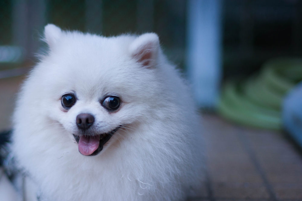
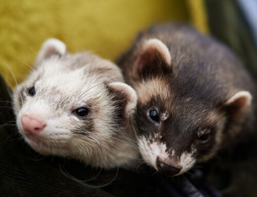
You’re playing with your dog and you notice something: one pupil looks noticeably larger than the other. Most owners in that moment have the same instinct, a quiet unease followed by the question of whether this is worth worrying about.
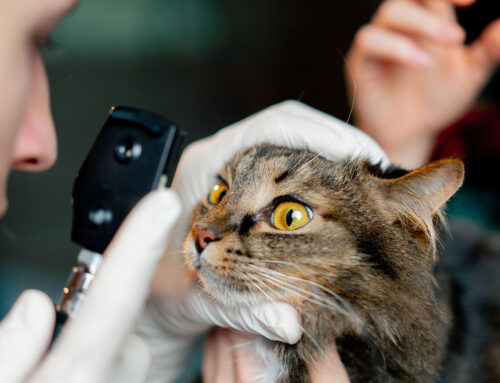
The answer is yes. Anisocoria, the medical term for unequal pupil size, is never a normal finding in dogs or cats, and it always warrants a prompt veterinary evaluation. The causes range from localized eye problems to neurological conditions affecting the brain, spinal cord, or the nerve pathways that control how pupils respond to light. Knowing which of those causes is responsible for what you’re seeing requires a systematic examination by a specialist who understands the full picture.
Veterinary Vision Center, led by board-certified ophthalmologist Dr. Pierce, is located inside University Veterinary Hospital in Shreveport, LA, and provides precisely that level of evaluation. The services at Veterinary Vision Center include advanced diagnostic testing and surgical therapies for the full range of ocular conditions that can produce anisocoria. Contact our practice if your pet’s pupils look uneven, especially if the change appears suddenly.
How Do Pupils Normally Work?
Pupils are the dark, circular openings in the center of the eye that control how much light reaches the retina. In bright conditions, the pupil constricts to a smaller size. In dim light, it dilates to allow more light in. This process is regulated by the autonomic nervous system, specifically two competing pathways: one that causes constriction and one that causes dilation. Both pathways run from the brain through the spinal cord and along nerves that reach the eye.
When anisocoria is present, one pupil is failing to respond the way it should. It may be too large, meaning it is not constricting appropriately in light, or it may be too small, meaning it is not dilating in the dark. Determining which pupil is the abnormal one is one of the first steps in diagnosis, and it matters enormously because the answer points toward completely different underlying causes.
A thorough ocular conditions evaluation at Veterinary Vision Center begins with exactly this assessment, using controlled lighting conditions and a detailed examination of both eyes together and individually.
What Causes Anisocoria in Dogs and Cats?
Eye-Specific Conditions That Affect Pupil Size
Many cases of anisocoria originate directly within the eye itself, where inflammation, pressure changes, structural damage, or abnormal tissue growth disrupt the normal mechanisms that control the pupil.
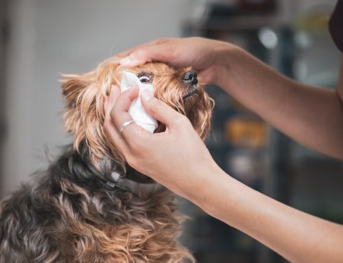
Uveitis, which is inflammation of the middle layer of the eye called the uvea, is one of the most common ocular causes. The inflammation can cause the pupil to constrict abnormally and in some cases become irregular in shape, sticking to surrounding structures through a process called synechia. It is painful, and affected eyes usually appear red, cloudy, or squinting.
Corneal ulcers are surface erosions on the clear front of the eye that cause pain and reflex tearing. When severe or deep, they can trigger a reflex constriction of the pupil on the affected side as a pain response. Cats in particular may develop corneal ulcers related to herpesvirus infection that produce exactly this presentation. The veterinary corneal ulcer services at Veterinary Vision Center address these cases with the diagnostic depth and treatment precision they require.
Glaucoma is a condition characterized by elevated pressure inside the eye, called intraocular pressure, that damages the optic nerve over time. It causes the affected pupil to appear dilated and unresponsive, and the eye may look enlarged, red, or hazy. Glaucoma is painful, progresses quickly, and causes irreversible vision loss without prompt intervention. It is one of the more urgent causes of anisocoria in dogs.
Lens dislocation, where the lens shifts out of its normal position, disrupts the anatomy of the eye in ways that directly affect the pupil. Some breeds are genetically predisposed, including Terrier breeds and Border Collies, and lens luxation can lead to secondary glaucoma if not treated promptly.
Eye tumors within or around the eye, whether primary ocular tumors or metastatic growths from cancer elsewhere in the body, can physically obstruct or distort normal pupil function. Iris melanomas in cats are a well-known example that can cause irregular or fixed pupils over time.
When the source of anisocoria appears to be within the eye itself, owners often notice accompanying signs: redness, cloudiness, squinting, increased tearing, or visible changes to the iris or cornea. The Veterinary Vision Center team evaluates all of these signs together to determine the source and the urgency.
Neurological Conditions That Cause Unequal Pupils
Because pupil size is controlled by the nervous system, conditions that affect the brain, spinal cord, or peripheral nerve pathways can all produce anisocoria without any visible abnormality in the eye itself. These cases are particularly important to identify because they often signal a broader systemic or neurological problem.
Horner’s syndrome is one of the most recognizable neurological causes of anisocoria in both dogs and cats. It results from disruption of the sympathetic nerve pathway, which runs from the hypothalamus in the brain, down through the cervical spinal cord, through the chest, and around the neck to the eye. The result is a cluster of signs on the affected side: a smaller pupil, a drooping upper eyelid, elevation of the third eyelid, and mild recession of the eyeball. Horner’s syndrome can result from neck or chest trauma, middle ear disease, a mass compressing the nerve, or in some cases it develops without an identifiable cause.
Brain tumors affecting areas that regulate the autonomic nervous system can cause pupil asymmetry, often alongside changes in behavior, coordination, consciousness, or appetite. The location of the lesion within the brain determines which eye is affected and what other signs appear alongside the anisocoria.
Strokes in dogs and cats, while less common than in humans, do occur and can produce sudden anisocoria as part of a broader neurological event. Affected pets may show a sudden head tilt, loss of balance, circling, or altered responsiveness in addition to the pupil asymmetry.
Optic nerve disorders, including inflammation, compression, or degeneration of the nerve that carries visual signals from the eye to the brain, can cause the pupil to respond abnormally to light even when the eye itself appears structurally normal. This is because the pupillary light reflex runs through the optic nerve before reaching the pupil-controlling pathways.
Head trauma can cause anisocoria (unequal pupil sizes) when the impact affects the brain or the nerves controlling the pupils. The brain pathways that regulate pupil size are sensitive to pressure and swelling, so even a bump to the head when playing can temporarily disrupt normal signaling. In more serious cases, like a pet being struck by a vehicle, bleeding or swelling inside the skull can compress the oculomotor nerve, causing one pupil to become fixed and dilated. Whether the injury seems minor or major, anisocoria after any head impact is a reason to contact your veterinarian promptly.
Neurological anisocoria often comes with other signs beyond the eyes: head tilt, stumbling or circling, facial drooping, one-sided weakness, or behavioral changes like disorientation or decreased interaction. These combinations always require urgent evaluation.
What Other Symptoms Should You Watch For?
Anisocoria does not always arrive alone. The combination of signs that accompany unequal pupils is one of the most useful diagnostic tools available to the veterinary team, because it narrows the list of possible causes significantly.
Watch for:
- Redness or cloudiness in one or both eyes
- Squinting or holding one eye shut
- Increased tearing or discharge
- The third eyelid (the tissue in the inner corner of the eye) becoming visible
- A drooping upper eyelid on one side
- Changes in vision: bumping into objects, hesitation in unfamiliar spaces, reduced response to movement
- Head tilt or leaning to one side
- Circling or loss of coordination
- Facial asymmetry or changes in the appearance of one side of the face
- Behavior changes: decreased appetite, lethargy, withdrawal, or unusual irritability
Sudden onset of any of these signs alongside anisocoria warrants immediate contact with Veterinary Vision Center. For after-hours emergencies, the practice can be reached at (318) 393-7033.
What Does the Diagnostic Process Look Like?
Evaluating anisocoria systematically is what allows us to distinguish between an eye-originating problem and a nervous system problem, which determines everything about what treatment is needed.
The examination typically includes:
- Detailed history. When did the owner first notice the asymmetry? Has it changed? Are there other signs? Any recent trauma, illness, or medication changes?
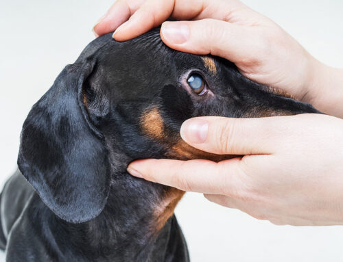
- Pupillary light reflex testing. A light is directed into each eye individually to assess whether the pupil constricts and whether that response is transmitted to the other eye as expected.
- Complete ophthalmic examination. This includes assessment of the cornea, anterior chamber, lens, iris, intraocular pressure measurement, and fundic examination of the retina and optic nerve.
- Neurological assessment. Gait, balance, reflexes, cranial nerve function, and response to visual stimuli all contribute information about whether the nervous system is involved.
- Additional diagnostics. Depending on initial findings, bloodwork, chest radiographs, advanced imaging such as MRI or CT, or referral for neurological consultation may be recommended to evaluate systemic causes or central nervous system involvement.
The goal is always a specific diagnosis rather than a working assumption, because effective treatment requires accurate identification of the source.
How Is Anisocoria Treated?
Treatment follows the diagnosis directly, and the range is broad because the causes are broad.
Eye-specific conditions are managed with targeted therapies: anti-inflammatory eye drops or systemic medications for uveitis, pressure-lowering medications or surgery for glaucoma, surgical intervention for lens luxation, and specialized care for corneal disease. Our practice at Veterinary Vision Center includes advanced surgical capabilities and a 95% success rate for cataract surgeries, reflecting the level of precision and experience brought to ocular procedures.
Neurological causes are managed in collaboration with the broader veterinary team, since conditions like brain tumors, strokes, or spinal cord lesions require systemic evaluation and treatment beyond ophthalmology alone. Horner’s syndrome with an identifiable underlying cause is treated by addressing that cause; idiopathic Horner’s syndrome, where no cause is found, often resolves on its own over weeks to months.
Follow-up rechecks are essential for tracking response to treatment, catching complications early, and adjusting therapies as the clinical picture evolves.
When Is Anisocoria an Emergency?
Some presentations of anisocoria require same-day or immediate care rather than a scheduled appointment.
Contact Veterinary Vision Center promptly or seek emergency care if:
- Anisocoria appeared suddenly, especially within the past 24 to 48 hours
- The eye appears red, cloudy, or visibly enlarged
- The pet is squinting intensely or pawing at the eye
- There are signs of pain: vocalizing, reluctance to be touched near the head, restlessness
- Vision appears affected: bumping into objects, reluctance to move, or inability to track movement
- Neurological signs are present: head tilt, loss of balance, circling, disorientation, collapse
- There has been any trauma to the head or eye area
Timing matters with many of these conditions. Glaucoma can cause permanent vision loss within hours of onset. Lens luxation becomes more complicated the longer it goes untreated. Brain lesions causing sudden anisocoria need rapid evaluation to guide management. Request an appointment at Veterinary Vision Center as soon as possible.
Frequently Asked Questions About Anisocoria in Pets
Can anisocoria in pets resolve on its own?
It depends entirely on the cause. Horner’s syndrome from an unknown cause sometimes resolves without treatment over several weeks. Most other causes, including glaucoma, uveitis, lens luxation, and neurological disease, will not resolve on their own and require treatment to prevent worsening or permanent damage. Waiting to see whether things improve is not advisable.
Is anisocoria painful?
It depends on the cause. Glaucoma, uveitis, and corneal ulcers are painful conditions. Horner’s syndrome and many neurological causes are not painful to the eye itself, though the underlying condition driving them may be. Signs of pain include squinting, pawing at the face, avoiding light, and behavioral changes.
How quickly do I need to act?
If anisocoria appeared suddenly, or if it is accompanied by redness, cloudiness, vision changes, or neurological signs, act the same day. For gradual onset without additional signs, contact the practice within 24 to 48 hours for guidance on urgency.
Protecting Your Pet’s Vision Starts With Noticing
Catching anisocoria early and responding promptly is one of the most meaningful things an owner can do for their pet’s eye health. The conditions that cause it range from very treatable when caught early to devastating when allowed to progress, and the difference often comes down to how quickly evaluation begins.
Dr. Pierce and the Veterinary Vision Center team approach every patient with the belief that your pet deserves the same quality of care you would want for yourself. World-class ophthalmology in a setting that treats families like family means you will leave knowing what is happening, why it matters, and what comes next. If your pet’s eyes are telling you something is wrong, trust that instinct and let the specialists take it from there- request an appointment today.
Leave A Comment