What allows you to see the breathtaking splendor of a sunset? The retina, an eye structure equal in beauty to a sunset—in the opinion of eye nerds, at least—plays an important role in vision. This 10-layered structure that lies against the back of the eye is responsible for changing an image into a nerve impulse, which travels up the optic nerve to the brain, and is then converted back to an image before being perceived by the brain, and therefore you. If the connection between the retina and the back of the eye is disrupted (i.e., a retinal detachment), the pet cannot see—akin to you unplugging your TV cable line and seeing a black, imageless screen.
Causes of retinal detachments in pets and horses
A variety of processes can be responsible for retinal detachments in pets, including:
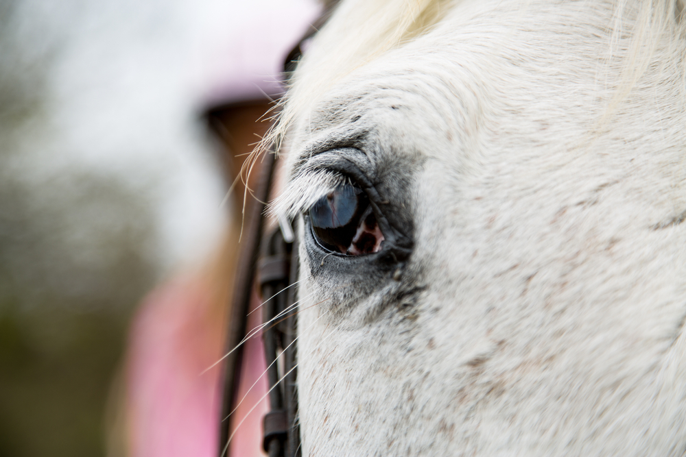
- Infections and inflammation — Fungal infections in dogs and cats, feline infectious peritonitis and feline leukemia virus in cats, and other infections can lead to retinal detachment in pets. Equine recurrent uveitis (ERU) commonly causes retinal detachment in horses.
- Congenital disorders — Some dogs or horses may suffer from breed-related retina or eye abnormalities that lead to retinal detachments.
- Trauma — Significant eye trauma can lead to partial or complete retinal detachment in horses, dogs, and cats.
- Eye surgery — Occasionally, retinal detachment occurs as a complication from cataract or lens removal surgery in dogs and horses.
- Hypertension (i.e., high blood pressure) — Hypertension is a common retinal detachment cause in older cats and typically occurs as a result of kidney disease or hyperthyroidism (i.e., increased thyroid hormone levels). Hypertension less commonly causes retinal detachment in dogs.
- Neoplasia (i.e., cancer) — Some cancer types that affect the back of the eye may lead to retinal detachment.
Retinal detachment signs in pets and horses
A retinal detachment may occur in one or both eyes, and typically leads to partial or complete blindness in the affected eye(s) because the vision pathway is disrupted. A pet or horse whose vision is impaired may be scared, reluctant to move, bump into objects, or have trouble navigating in different light levels. Horses who become blind in one eye may spook at sounds or sensations on their blind side, but pets who are blind in one eye often compensate well and may show few signs until the other eye also becomes blind. Affected animals may have a dilated pupil or a reddish haze to the inside of the eye from intraocular bleeding (i.e., hyphema).
Retinal detachment diagnosis in pets and horses
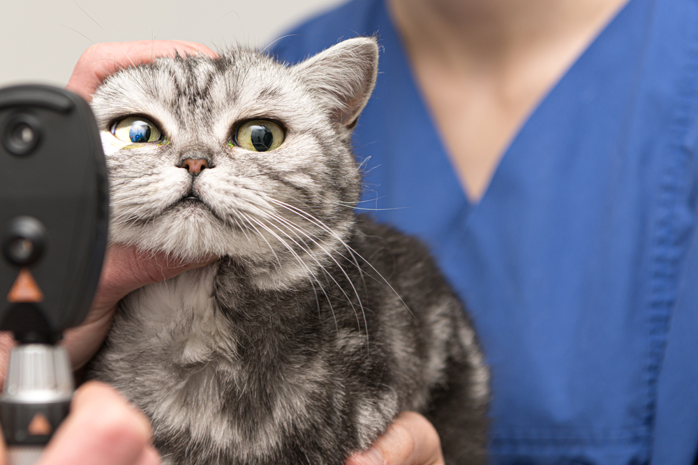
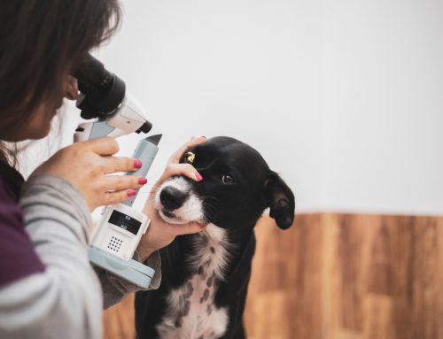
Dr. Pierce will perform a complete ophthalmic examination, which involves measuring the eye pressures, viewing the eye structures with a handheld microscope, visualizing the retina with a headlamp and a set of special lenses, and other tests. He may also watch your pet navigate an unfamiliar maze of objects in different light conditions, shine a bright light in your pet’s eye to see if they blink or pull back their head (i.e., dazzle test) or they blink or pull back if he moves his hand toward their face (i.e., menace test), and assess the eye’s light response (i.e., pupillary light reflex). To better characterize or diagnose a retinal detachment and evaluate other eye structures, Dr. Pierce may also perform an eye ultrasound. If high blood pressure or systemic disease are suspected to cause the detached retina, he may measure your pet’s blood pressure or test for fungal, viral, or bacterial diseases.
Management of retinal detachments in pets and horses
Retinal detachment management depends on the reason for the detachment and whether the detachment is partial or complete. Pets with hypertension must be treated with blood pressure- lowering medications to control their blood pressure. Anti-inflammatory medications or disease-specific treatments may be used to address infectious or inflammatory diseases. In some cases, Dr. Pierce may perform a retinopexy, which is a surgical procedure used to tack the retina down around a partial detachment to prevent the detached area from increasing, or to preventively tack down the retina in a pet who has one detached eye and is at high risk of detachment in the other eye.
Long-term outlook for pets and horses with retinal detachment
Some pets with retinal detachments will get their sight back once the underlying condition is managed, while others will remain blind. Pets suffering a hypertension-induced retinal detachment may regain vision over a period of six to eight weeks as their blood pressure is managed, unless the retina damage is irreversible. The good news is that blind pets usually adapt well and have a good quality of life, especially with a dedicated owner and a relatively unchanging environment. A newly blind horse can be more challenging, but these resources may help you and your horse make a smooth transition.
The sooner a retinal detachment is diagnosed and managed, the better the chance that the retina will reattach or surgery will be successful and preserve some vision. If you have any concerns about your pet’s or horse’s vision, don’t hesitate to contact us at Veterinary Vision Center—after all, vision is our middle name.
Leave A Comment