
Cats are prone to all sorts of eye mishaps, and an accidental scratch can lead to a corneal ulcer, and possibly a corneal sequestrum. Corneal sequestra develop fairly often following feline ocular injuries, although cat owners are rarely familiar with this condition until they notice an odd spot on their cat’s eye and take them to their family veterinarian. If you have a curious cat who gets into mischief and may scratch their eye, or you see an abnormality on their eye surface, you need to know about corneal sequestra, and the signs.
What is a feline corneal sequestrum?
Your cat’s cornea is the transparent surface at the front of their eyeball that allows light to enter. A corneal sequestrum is an area of dead (i.e., necrotic) corneal tissue, which appears as a pigmented spot, ranging from tan to black, on the normally clear corneal surface. A corneal sequestrum that forms acts as a foreign body on the cornea, and stimulates inflammation and abnormal blood vessel development.
What causes a feline corneal sequestrum?
A corneal sequestrum typically results from a corneal ulcer, which is a scratch or abrasion on the corneal surface. Corneal ulcers often result from eye injuries, such as a scratch caused by another pet, or when your pet brushes past outdoor plants. The cornea normally heals quickly, despite lacking blood supply, and most corneal ulcers resolve without complication. However, an ulcer that is deep, becomes infected, or does not receive proper treatment may persist, and develop into a corneal sequestrum. Dead tissue fills the corneal defect, like a scab on the skin, and further interferes with healing.
Any cat can develop a corneal sequestrum, but the condition is more prevalent in brachycephalic breeds, such as Persians and Himalayans. Since their eyes are closer to the front of their face, and their short muzzles offer little protection, these breeds are more likely to injure their cornea. Cats with chronic feline herpesvirus infection, also known as feline viral rhinotracheitis, also are more likely to develop corneal ulcers, and corneal sequestra.
What are corneal sequestrum signs in cats?
A corneal sequestrum causes ocular inflammation, and mild to severe discomfort in affected cats, who may show signs that include:
- A colored spot on the cornea
- A cloudy cornea
- Blood vessels on the cornea
- Tearing
- Squinting
- Mucoid ocular discharge
- Third eyelid elevation
- Ocular redness
- Eyelid swelling
If you notice any ocular inflammation signs in your cat, schedule an evaluation with your family veterinarian or our Veterinary Vision Center team immediately. While many eye conditions heal without complications, some can cause blindness without prompt treatment.
How is a corneal sequestrum diagnosed in cats?
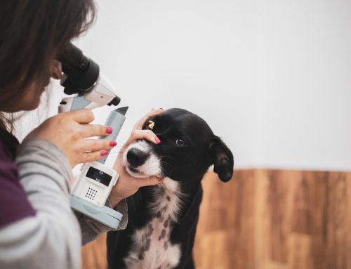
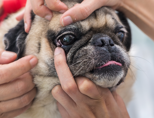
Your family veterinarian can typically diagnose a corneal sequestrum during a thorough ocular exam. They may also stain your cat’s cornea to evaluate the sequestrum and surrounding tissue. Corneal staining involves applying a few drops of bright yellow fluorescein to your cat’s corneal surface. Intact corneal tissue does not take up the stain, but damaged corneal tissue, including ulcerated or necrosed areas, will absorb the stain and fluoresce under a black light.
If your family veterinarian diagnoses a corneal sequestrum, they may refer your cat to a board-certified veterinary ophthalmologist for further diagnostic tests and management, since the condition can progress and lead to further complications if not treated appropriately.
Can a corneal sequestrum affect my cat’s vision?
Since a corneal sequestrum is pigmented, rather than transparent, it can partially obstruct your cat’s vision. The degree of impairment will depend on the sequestrum’s severity and location, as well as additional corneal damage, such as edema. A thorough work-up will include assessing your cat’s vision, as the initial trauma that led to the corneal sequestrum may have caused additional damage that could interfere with vision, or cause blindness.
How is a feline corneal sequestrum treated?

Every cat with a corneal sequestrum should be evaluated by a board-certified veterinary ophthalmologist, such as Veterinary Vision Center’s Dr. Kenneth Pierce, who will determine the best treatment plan. Some cats with superficial corneal sequestra may do well with medical management, but many cases require surgery for full resolution. Cats who are uncomfortable and display signs such as tearing, squinting, and redness need surgical treatment for more immediate relief.
Corneal sequestrum surgical treatment involves performing a keratectomy (i.e., removing the top layers of affected cornea). Dr. Pierce will then make a conjunctival flap from tissue beneath the cat’s eyelid, and suture it over the defect to aid healing. The cornea lacks blood supply, and the conjunctival tissue’s blood vessels will supply the affected area with nutrient-rich blood that enhances healing. Dr. Pierce may also temporarily suture the cat’s eyelid closed to further protect the healing cornea. After the conjunctival graft permanently adheres to the cornea, Dr. Pierce will sever its connection, cutting off blood supply that is no longer needed. Over time, the conjunctival tissue that remains over the healed ulcer will lose its pink appearance and become a scar that may mildly impede vision, but the cat’s eye will no longer cause discomfort or be at risk for blindness or rupture.
If you notice a colored spot on your cat’s eye, or signs of ocular inflammation, act quickly to preserve their vision. Our Veterinary Vision Center team is here to help with any ocular condition your pet may develop—contact us.

Leave A Comment