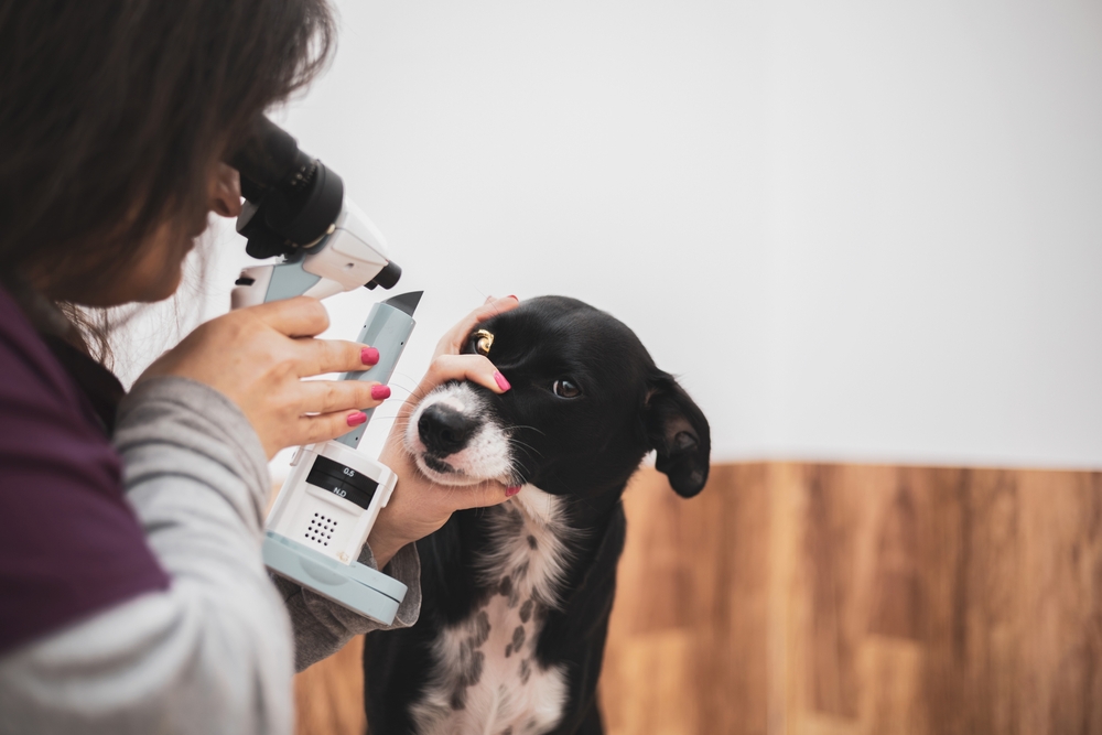
Testing a dog or cat’s vision requires specialized equipment and techniques, because patients who can’t read an eye chart call for a different approach entirely. Veterinary ophthalmologists use a battery of behavioral and instrument-based tests to evaluate not just whether an animal can see, but how well each part of the visual system is functioning, from the tear film and cornea to the retina and the neural pathways that carry signals to the brain. Early detection of conditions like progressive retinal atrophy, glaucoma, or cataract formation can make a meaningful difference in outcomes, since some causes of vision loss are manageable or even reversible when caught early.
Veterinary Vision Center in Shreveport is a referral ophthalmology practice led by a board-certified veterinary ophthalmologist, offering the full range of advanced ophthalmic diagnostics, including electroretinography, tonometry, gonioscopy, ocular ultrasonography, and Schirmer tear testing. Our ophthalmic services are designed for cases that go beyond what a general practice can evaluate in-house, and we work closely with referring veterinarians to ensure continuity of care. If your veterinarian has recommended an ophthalmology consultation, or if you have noticed changes in your pet’s eyes or behavior that concern you, reach out to us to arrange an appointment.
Pet Vision Testing Quick Guide
- Two kinds of testing: vision testing uses behavioral observations (menace response, pupillary reflexes, obstacle navigation) plus instrument-based testing.
- Specialty tools see deeper: electroretinography, ocular ultrasound, gonioscopy, and tonometry provide depth that general practice exams cannot match.
- Pets hide loss well: subtle behavioral changes often signal vision problems before you notice, because pets compensate for gradual loss.
- Early detection matters: it meaningfully changes outcomes for treatable conditions like glaucoma, dry eye, and cataracts.
How Does Animal Vision Work?
Dogs and cats see the world on different terms than people do, and the basic eye structure and function of a pet eye explains a lot of why testing it takes specialized tools. The key differences:
| Visual feature | How dogs and cats compare to humans |
| Color perception | A more limited range (mainly blue and yellow); color is not their primary cue |
| Night vision | Far more efficient in low light, thanks to a reflective tapetum lucidum |
| Field of view | Wider (most dogs ~240 degrees vs ~180 in humans) because eyes sit more laterally |
| Visual acuity | Lower sharpness than humans, but exceptional movement detection |
| Pupil shape | Cats have vertical slit pupils that adjust rapidly; dogs have round pupils |
The eye structures work together to process light: cornea, lens, vitreous, retina, and optic nerve each play roles, with the visual cortex of the brain interpreting the signals. Understanding normal function helps identify when something goes wrong, since each structure can fail in characteristic ways that point toward specific diagnoses.
Vision is one of several senses pets rely on alongside smell, hearing, and touch, and they lean harder on the others when sight slips. That cross-compensation is part of why early vision changes go unnoticed at home for so long.
How Do Veterinarians Test Pet Vision?
The diagnostic process moves from general observations to specialized testing.
Initial Eye Examination Techniques
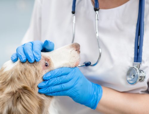
The foundational eye exam includes:
- Visual inspection: external structures (eyelids, lashes, conjunctiva, cornea, anterior chamber) under good light and magnification. This first look catches problems like inward-rolling eyelids, extra lashes rubbing the cornea, redness, discharge, cloudiness, or a difference between the two eyes. Much of what affects vision starts on the surface, so a careful external survey often points the rest of the exam in the right direction.
- Pupillary light reflex: tests the neurological pathway from retina to brain to pupil. A normal response indicates the basic visual pathway is intact. We shine a light into one eye and watch both pupils constrict, which confirms the retina is sensing light and the nerves are carrying that signal. On its own it does not prove conscious sight, since a pet can have intact reflexes yet still be blind from a problem higher in the brain, which is why we pair it with other tests.
- Menace response: a hand motion toward the eye should produce a blink, testing visual perception plus reflex coordination. We make a quick gesture toward the eye, careful not to create a breeze or touch the whiskers, since either could trigger a blink by feel rather than sight. A reliable blink tells us the pet both sees the motion and can coordinate a protective response, though very young puppies and kittens may not have developed it yet.
- Tracking movements: a pet that visually follows a thrown cotton ball (silent, so they cannot track by sound) is responding to vision. Cotton balls are used precisely because they fall without a sound or scent to follow, so any tracking has to come from sight. Watching whether the eyes and head follow the object, and how smoothly they do it, gives a real-world read on functional vision that instruments alone cannot.
- Dazzle reflex: a bright light should cause a squint or partial blink. This is a deeper, more primitive reflex than the menace response, routed through the brainstem, so it can remain present even in a pet that has lost conscious vision. We use it as one more clue about where along the visual pathway a problem sits, which is especially helpful in eyes too cloudy to examine directly.
Each step of a physical eye exam targets a specific structure or function. Pets typically tolerate exam techniques well, though some elements require specialized lighting (a dim room) or briefly dilated pupils for full evaluation.
Specialized Diagnostic Tests for Eye Health
Advanced testing methods provide more thorough assessment. Common ocular tests include:
- Tonometry: measures intraocular pressure to screen for glaucoma. A brief, painless test using a handheld device. After a drop of numbing anesthetic, the instrument gently touches the surface of the eye and reads the internal pressure in seconds. Catching elevated pressure early matters enormously, since glaucoma can destroy vision within hours to days, and unusually low pressure can instead point toward inflammation inside the eye.
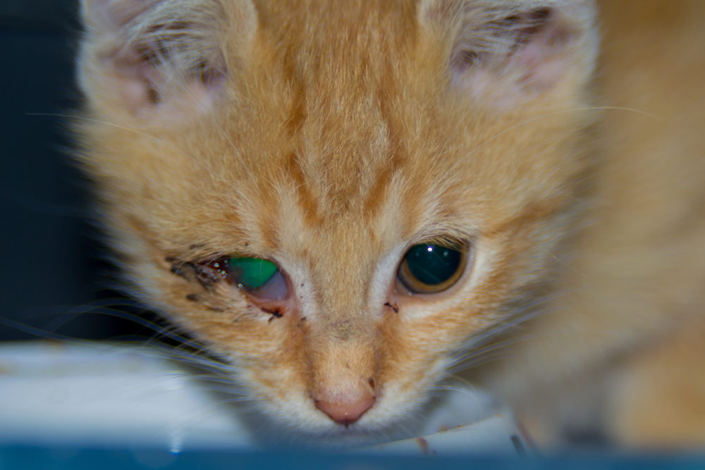
- Fluorescein stain: a green dye applied to the eye reveals corneal damage. Critical for diagnosing ulcers. The dye clings to areas where the cornea’s protective surface is broken and glows under a blue light, mapping out scratches and ulcers that are otherwise invisible. It also helps us judge how deep an ulcer runs and whether the tear drainage system into the nose is working normally.
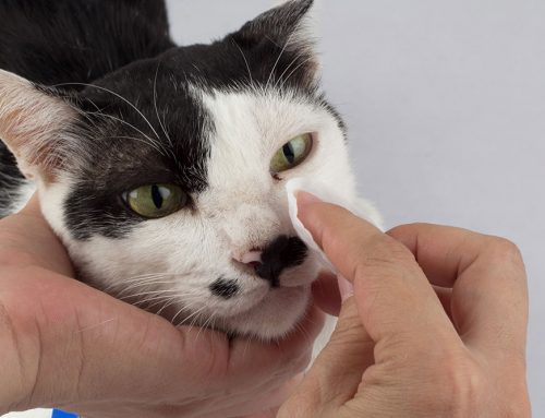
- Schirmer tear test: a small paper strip measures tear production over 60 seconds. Diagnoses dry eye. The strip tucks gently inside the lower lid and wicks up tears, and the distance the moisture travels shows whether the eye is making enough of them. Dry eye is common, uncomfortable, and very treatable, but left undiagnosed it leads to chronic infections, pigment buildup, and vision loss, so this quick measurement earns its place.
- Tear film assessment and osmolarity: advanced tear evaluation beyond simple production. Healthy tears are about quality as much as quantity, with oily, watery, and mucus layers that all have to balance. These measurements look at how stable and how concentrated the tear film is, which helps explain ongoing irritation in pets whose basic tear production looks normal on paper.
- Gonioscopy: evaluates the drainage angle of the eye for glaucoma assessment. Using a special lens placed on the eye, we examine the tiny angle where fluid normally drains out, since a narrow or malformed angle predisposes many breeds to glaucoma. Checking it can flag a high-risk eye before pressure ever climbs, and it guides how we protect the second eye when glaucoma has already struck the first.
- Slit lamp biomicroscopy: magnified, illuminated examination of anterior eye structures. This instrument throws a thin, bright slice of light through the eye and magnifies it, letting us study the cornea, lens, and front chamber in fine detail. It reveals subtle changes like early cataracts, low-grade inflammation, or tiny corneal deposits that a routine light and the naked eye would miss.
- Fundic examination: direct or indirect ophthalmoscopy to evaluate the retina and optic nerve. By looking through the pupil, often after dilating it, we view the retina and optic nerve at the back of the eye, where vision actually begins and where many sight-threatening diseases hide. Changes here can reveal retinal degeneration, detachment, high blood pressure damage, and sometimes signs of disease elsewhere in the body.
- Electroretinography (ERG): measures the electrical response of the retina to light. Critical for assessing retinal function, particularly before cataract surgery to confirm the retina works adequately to benefit from the procedure. The retina produces a tiny electrical signal when light strikes it, and the ERG records that signal to confirm the retina is alive and functioning even when we cannot see it directly. This is essential before cataract surgery, since clearing a cloudy lens does no good if the retina behind it has failed, and it is also how conditions like progressive retinal atrophy are confirmed.
Each test answers specific questions about specific parts of the visual system, and the combination provides a complete picture.
Diagnostic Imaging for Eye Conditions
When direct visualization is limited by cloudiness, inflammation, or other obstacles, imaging fills the gap. Ocular ultrasonography is the most common imaging tool in veterinary ophthalmology:
- Behind cataracts: when the lens is opaque, ultrasound assesses whether the retina is normal before considering cataract surgery. A mature cataract blocks our view of the back of the eye entirely, so ultrasound becomes the only way to confirm the retina is attached and healthy underneath. That answer directly shapes whether surgery is worth pursuing, since a detached retina would mean the procedure could not restore sight.
- Intraocular masses: identifies tumors that would not be visible through external exam. Sound waves can outline a growth inside the eye, showing its size, location, and relationship to nearby structures. That information helps distinguish a harmless cyst from a tumor and guides whether monitoring, biopsy, or removal is the right next step.
- Retinal detachment: confirms suspected detachment. When a pet loses vision suddenly and the retina cannot be seen clearly, ultrasound shows whether it has pulled away from the back of the eye. Confirming a detachment quickly matters, because some can be reattached when the underlying cause, such as high blood pressure, is addressed in time.
- Vitreous abnormalities: bleeding, debris, or other changes within the eye. The vitreous is the clear gel filling the back of the eye, and ultrasound reveals blood, inflammatory debris, or strands floating within it. These findings often point toward an underlying problem, such as trauma, a clotting disorder, or inflammation, that needs its own workup.
- Trauma assessment: when the eye is too swollen or painful for detailed internal examination. After an injury, swelling and pain can make a hands-on internal exam impossible, but ultrasound sees through that to assess the lens, retina, and eye wall. It helps us determine how much damage the eye has sustained and whether it can be saved, all without causing further discomfort.
The procedure is non-invasive and well-tolerated, usually requiring only mild sedation or sometimes none at all. Other advanced imaging (CT, MRI) is used for cases extending to the orbit, optic pathway, or brain.
Frequently Asked Questions About Pet Vision and Eye Health
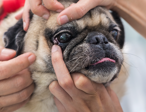
How can I tell if my pet has lost vision in one eye?
Cover each eye briefly during play or feeding. If your pet has trouble tracking thrown objects or navigating with one eye covered but does fine with the other covered, asymmetric vision is likely. A specialty ophthalmic exam can quantify and characterize what is happening.
Are vision tests painful or stressful for pets?
Most are not. The exam relies on observation, handheld instruments, and gentle techniques. Pets that need sedation for advanced tests (ocular ultrasound, certain surgical evaluations) tolerate the sedation well.
How often should my pet have a vision screening?
For most pets, an annual wellness exam includes a basic eye assessment. Specialty ophthalmology screening makes sense for senior pets, breeds with known eye disease predispositions, pets with diabetes or other systemic disease affecting the eyes, and any pet showing concerning signs.
Can my pet see in color?
They can, but less vividly than humans. Dogs primarily see in shades of blue and yellow, and cats see similarly. Their color vision is functional but not as rich as human color perception.
Catching Vision Trouble Early Is the Whole Point
Routine eye screening and early intervention are how vision and comfort get preserved through every life stage. Plenty of eye conditions are manageable or even treatable when caught early, and the screening that catches them does not need dramatic symptoms to be worth doing.
If your veterinarian has recommended an ophthalmology consultation, or if you have noticed concerning vision changes in your pet, request an appointment or reach out to us and our team will help.
Leave A Comment