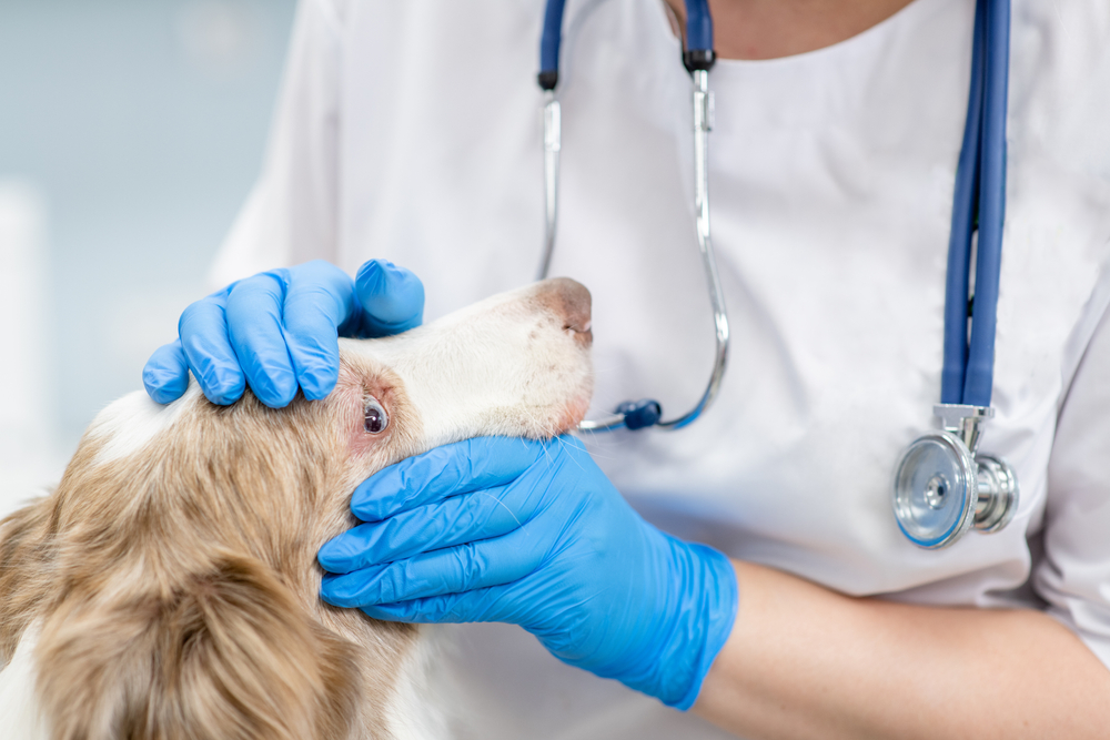
Not all changes to a senior pet’s eyes turn out to be normal aging, and the changes that do threaten vision are often the most manageable when they are caught early. A bluish haze in the lens is usually lenticular sclerosis, a normal aging change with minimal effect on functional vision, but a white or opaque lens may indicate a cataract that can cause significant vision loss and may be surgically treatable. Glaucoma, dry eye, and retinal degeneration become more common as pets age, and most are silent in the early stages, not causing the obvious distress you might expect given how uncomfortable they can become. Routine vision screening in senior pets is how these conditions get caught at a stage where there is still something meaningful to do.
Veterinary Vision Center in Shreveport is a veterinary ophthalmology specialty practice led by a board-certified veterinary ophthalmologist, and senior vision screening is a core part of what we do. Our diagnostic capabilities include electroretinography, ocular ultrasonography, tonometry, gonioscopy, and tear film assessment, the same tools used to evaluate age-related eye disease in the earliest and most treatable stages. Reach out to us to schedule an appointment for your senior pet.
Senior Eye Screening Quick Guide
- Two look-alikes, different conditions: lenticular sclerosis (bluish haze) and cataracts (white opacity) look similar but only an exam reliably distinguishes them.
- Glaucoma is fast: it can cause permanent blindness within hours of pressure elevation, so pressure checks at wellness exams catch it early.
- Pets hide vision loss well: subtle behavioral changes often signal vision problems before owners realize.
- Specialty tools see more: electroretinography, ocular ultrasound, and gonioscopy provide diagnostic depth that general practice exams cannot replicate.
Why Does Vision Screening Matter for Senior Pets?
As pets age, the lens, retina, cornea, and other eye structures undergo natural changes. Some changes are normal and benign; others can progress to vision impairment or blindness. The distinction matters because the management differs entirely.
Preventive testing builds the baseline that makes change detection meaningful over time. Regular screening allows differentiation between harmless aging and conditions requiring intervention, which improves outcomes and maintains a pet’s independence and confidence. A pet who has lost partial vision adapts well to a familiar environment, but the underlying condition and its trajectory is the question worth answering.
Pet owners often delay vision screening because nothing seems obviously wrong. The reality is that:
- Slow progression hides disease: most senior eye diseases advance gradually enough that day-to-day observation does not catch them.
- Pets compensate: they adapt smoothly to slow change, masking vision loss in ways that fool the people who live with them.
- Look-alikes diverge: many conditions that look identical externally have completely different prognoses.
- Early action pays off: intervention preserves more vision and prevents secondary complications.
The strongest pet outcomes come from baseline screening followed by periodic rechecks, with specialty involvement when something needs deeper evaluation.
Which Eye Conditions Are Common in Senior Pets?
A range of conditions becomes more common with age, from benign changes that require only monitoring to serious diseases requiring active treatment.
Distinguishing Cloudiness in the Lens
Two common causes of cloudy eyes in older pets look similar at first glance but are completely different:
- Nuclear sclerosis: a normal aging change where the lens hardens and develops a bluish-gray appearance. Vision is generally preserved; the pet sees through the change rather than around it. No treatment is needed beyond monitoring.
- Cataracts: lens opacities that block light from reaching the retina. Mature cataracts cause functional blindness in the affected eye and can trigger inflammation (uveitis) and secondary glaucoma if untreated.
The cataracts vs nuclear sclerosis distinction matters because nuclear sclerosis requires monitoring only, while cataracts may benefit from surgical removal that restores vision in carefully selected patients. A dog who develops sudden cataracts should be screened for diabetes; rapid-onset bilateral cataracts in a previously healthy dog often turn out to be the first sign of diabetes mellitus, and untreated cataracts can cause secondary complications. A thorough ophthalmic exam reliably distinguishes the two and guides appropriate management.
Glaucoma and Intraocular Pressure
Glaucoma is elevated pressure inside the eye, and it is genuinely an emergency when acute. Untreated acute glaucoma can cause permanent blindness within hours to days.
Signs include redness, cloudiness (the eye may look bluish or hazy), enlargement of the eye, pain (squinting, pawing at the face, lethargy), and sometimes a dilated pupil. Senior pets are at higher risk, and certain breeds (Cocker Spaniels, Basset Hounds, Bostons, Beagles, and others) are predisposed to primary glaucoma.
Routine pressure checks during senior wellness visits and ophthalmology exams catch glaucoma early. Tonometry is a brief, painless test that measures intraocular pressure in seconds. For at-risk breeds, periodic screening can identify pressure elevation before clinical signs appear, allowing medical management to preserve vision and comfort.
Retinal Diseases and Degeneration
The retina is the light-sensitive tissue at the back of the eye, and several conditions affect it specifically in senior pets:
- Progressive retinal atrophy (PRA): inherited degeneration of the retina that eventually causes blindness. More commonly identified in middle-aged pets, but progression continues into senior years.
- Sudden acquired retinal degeneration syndrome (SARDS): causes acute blindness, often within days, in middle-aged to older dogs. Cause incompletely understood; vision loss usually irreversible.
- Hypertensive retinopathy: high blood pressure damages the retina, sometimes causing sudden vision loss. Often associated with systemic hypertension, which is itself often secondary to kidney disease, hyperthyroidism (cats), or heart disease.
- Retinal detachment: sudden vision loss with multiple potential causes including trauma, hypertension, inflammation, and certain systemic diseases.
Fundic examination (looking at the back of the eye) and blood pressure screening are core parts of detecting these conditions. The treatable categories (hypertensive retinopathy especially) benefit from early intervention; the irreversible ones still benefit from accurate diagnosis so families can plan and adjust home environments.
Dry Eye and Tear Production Changes
Dry eye (keratoconjunctivitis sicca, KCS) is reduced tear production leading to chronic eye irritation. Senior pets may develop dry eye due to:
- Immune-mediated destruction of tear glands (most common cause).
- Medications that can suppress tear production.
- Age-related gland changes.
- Certain systemic diseases.
Signs include thick, ropey discharge, redness, squinting, and cloudiness from chronic surface inflammation. Schirmer tear testing measures tear production in 60 seconds and identifies dry eye reliably. Treatment is usually lifelong: cyclosporine or tacrolimus drops to stimulate tear production, plus lubricating drops or ointments to supplement what is missing. With consistent treatment, most cases stay well-controlled and vision is preserved.
Eyelid Tumors and Growths
Eyelid masses are common in senior pets, and eyelid tumors vary widely:
- Dogs: more often benign (meibomian gland adenomas, papillomas). Surgical removal prevents corneal irritation as they grow and confirms histology.
- Cats: more likely to be malignant, so biopsy and timely surgical planning matter more.
Mass evaluation considers size, growth rate, location, and corneal contact. Even benign masses warrant removal if they irritate the eye, grow rapidly, or change in appearance.
Breed-Specific and Inflammatory Eye Conditions
Pannus (chronic superficial keratitis) is an immune-mediated condition affecting the cornea, primarily in middle-aged and senior German Shepherds and a few related breeds. UV exposure intensifies pannus, which makes the condition particularly relevant in sunny climates like Louisiana. Most cases respond well to immunomodulatory eye drops with consistent treatment, and protective eyewear can meaningfully reduce flares.
Other immune-mediated and inflammatory eye conditions can develop or worsen with age. The specialty diagnostic tools available at an ophthalmology practice (gonioscopy, advanced imaging, ocular ultrasound) provide depth that general practice exams cannot match.
What Are the Signs of Vision Problems at Home?
Pets are quietly good at compensating for gradual vision loss, which is exactly what makes the subtle changes easy to miss. Behavioral changes to watch for:
- Hesitation on stairs, especially going down.
- Bumping into objects, especially after rearranging furniture.
- Reluctance to navigate in dim light.
- Increased startle responses when approached.
- Reduced confidence in unfamiliar environments.
- Following walls or familiar paths in the home.
- Difficulty finding food or water bowls.
- Changes in eye appearance: cloudiness, color changes, redness.
- Squinting or holding an eye closed.
- Pawing at the face.
- Increased tearing or discharge.
Monitor each eye individually; pets can compensate completely with one good eye while losing vision in the other.
Acute blindness (sudden vision loss over hours to days) is always urgent and warrants immediate evaluation.
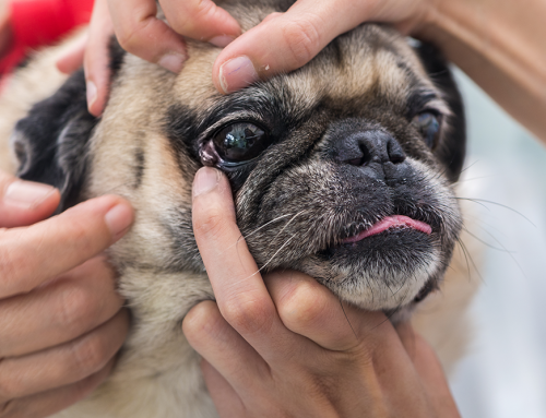
What Happens During a Senior Vision Screening?
A senior vision screening combines a thorough ophthalmic examination with additional diagnostic testing when needed.
The Comprehensive Ophthalmic Examination
A specialty ophthalmic exam works through these components:
| Exam component | What it screens for |
| External evaluation | Eyelids, lashes, surrounding structures |
| Schirmer tear test | Dry eye (tear production) |
| Tonometry | Glaucoma (intraocular pressure) |
| Slit lamp biomicroscopy | Cornea and lens |
| Pupillary light reflex | Retinal and neurological function |
| Fundic examination | Retina and optic nerve |
| Fluorescein stain | Corneal ulcers |
Dilation drops may be used to help visualize internal structures, tear film assessment goes beyond basic tear quantity, and most elements of the exam are non-invasive and well-tolerated. A thorough first visit typically takes 30 to 60 minutes.
Additional Diagnostic Testing When Needed
Some conditions require further diagnostics:
| Advanced test | When it is used |
| Ocular ultrasonography | Examining structures behind cataracts; identifying intraocular masses |
| Electroretinography (ERG) | Assessing retinal function, especially before cataract surgery |
| Gonioscopy | Evaluating the drainage angle for glaucoma assessment |
| Blood pressure measurement | Suspected hypertensive retinopathy |
| Bloodwork | Systemic disease contributing to ocular signs |
Our specialty practice has the equipment for all of these tests in one place, which streamlines the workup and avoids referrals to multiple facilities.
How Are Senior Eye Conditions Treated?
Treatment approaches range from ongoing medical management to surgical intervention, depending on the condition and severity.
Medical Management and Monitoring
Many conditions are managed with medications, lifestyle modifications, and regular monitoring, with goals to:
- Preserve remaining vision.
- Maintain comfort.
- Prevent progression.
- Catch complications early.
Practical guidance includes proper eye medication administration, completing prescribed treatment courses, and adjusting home environments for pets with vision changes. Owner education is a core part of the management plan; eye drops administered incorrectly do not deliver the intended effect.
Surgical Interventions When Appropriate
Some conditions benefit from surgical treatment:
- Cataract surgery (phacoemulsification): restores vision in selected patients. Our practice has a 95% success rate for appropriately selected cases, and pre-surgical ERG confirms retinal function is adequate to benefit from removal.
- Eyelid tumor excision: removes masses that irritate the eye or warrant histology.
- Glaucoma procedures: various options for reducing pressure or managing painful blind eyes.
- Corneal ulcer repair: surgical treatment for deep or non-healing ulcers.
- Enucleation (eye removal): in select cases where the eye cannot be saved and is causing intractable pain. Most pets adjust well to having one eye within weeks.
We discuss surgical options, timing, risks, and expected outcomes specific to each patient, and coordinate with referring veterinarians on perioperative care.
How Can I Support a Pet Losing Vision?
When vision loss occurs, practical adaptations help pets maintain confidence and quality of life.
Adapting the Home Environment
These tips help pets with vision loss navigate confidently:
- Keep furniture, food, and water bowls in consistent locations.
- Avoid rearranging rooms.
- Add textured mats or rugs to define pathways.
- Increase lighting in dim areas.
- Use verbal cues to announce your approach.
- Use scent markers (a specific essential oil dabbed at locations) sparingly to mark important areas.
- Block access to stairs with baby gates if balance is also affected.
- Cover sharp corners on furniture.
- Keep walkways clear of obstacles.
Most blind and low-vision pets settle into a remarkable rhythm once the home is set up around them.
Maintaining Enrichment and Connection
Visually impaired pets thrive on:
- Scent games: hide treats around familiar areas.
- Auditory toys: squeakers, crinkle materials.
- Tactile play: textured toys, snuffle mats.
- Consistent daily routines.
- Verbal interaction and reassurance.
- Walks on familiar paths.
Blind and low-vision pets often live happy, full lives, and families regularly say they did not realize how complete the adaptation had become until they stepped back and watched their pet navigate the house with confidence.

Frequently Asked Questions About Senior Pet Vision Health
My older dog’s eyes look cloudy. Is it cataracts?
Maybe, maybe not. The bluish-gray haze of nuclear sclerosis (normal aging) looks similar to early cataracts but is benign. Only an ophthalmic exam reliably distinguishes the two. If your dog is also showing increased thirst, urination, or weight loss, cataracts should prompt diabetes screening.
How often should my senior pet have a vision screening?
Annually for most senior pets, and more often for at-risk breeds or pets with known eye conditions. For specialty ophthalmology, baseline screening around age 7 to 8 with rechecks every 1 to 2 years is reasonable, and more frequent for pets with diagnosed conditions.
My cat doesn’t seem bothered by anything. Does she really need a vision check?
Cats are exceptionally good at hiding vision changes. Many cats with significant vision loss act entirely normally in familiar environments. A baseline screening identifies what is changing and lets you anticipate adaptations.
Will my dog need anesthesia for the eye exam?
Most ophthalmic exams are done awake with gentle handling. Some procedures (ocular ultrasound, certain advanced tests, surgical procedures) require sedation or anesthesia. We discuss what is needed and the anesthetic plan in advance.
What if my pet goes blind suddenly?
Treat sudden vision loss as urgent. Some causes are treatable if caught immediately (retinal detachment, hypertensive retinopathy with prompt blood pressure control), while others are irreversible (SARDS, severe optic neuritis). Either way, a prompt evaluation determines the cause and the right next steps.
Keeping a Senior Pet Seeing Comfortably
Regular vision screening is one of the quieter cornerstones of senior pet care. Early detection of the treatable conditions, like glaucoma, dry eye, and hypertensive retinopathy, is what separates preserved vision from irreversible loss, and accurate diagnosis lets families plan and adapt confidently when changes are permanent. Either outcome leaves a pet more comfortable and a family less surprised, which is what the screening is really for.
If your senior dog or cat would benefit from a vision screening, or if you have noticed changes in their eyes or behavior, request an appointment or reach out to us and our team will help.
Leave A Comment